5O0U | pdb_00005o0u

Crystal structure of tarantula venom peptide Protoxin-II


Experimental Data Snapshot

  • Method: X-RAY DIFFRACTION
  • Resolution: 0.99 Å
  • R-Value Free: 
    0.140 (Depositor), 0.154 (DCC) 
  • R-Value Work: 
    0.126 (Depositor) 
  • R-Value Observed: 
    0.127 (Depositor) 

wwPDB Validation 3D Report Full Report

Validation slider image for 5O0U

This is version 1.2 of the entry. See complete history

Literature

The Role of Disulfide Bond Replacements in Analogues of the Tarantula Toxin ProTx-II and Their Effects on Inhibition of the Voltage-Gated Sodium Ion Channel Nav1.7.

Wright, Z.V.F.McCarthy, S.Dickman, R.Reyes, F.E.Sanchez-Martinez, S.Cryar, A.Kilford, I.Hall, A.Takle, A.K.Topf, M.Gonen, T.Thalassinos, K.Tabor, A.B.

(2017) J Am Chem Soc 139: 13063-13075

  • DOI: https://doi.org/10.1021/jacs.7b06506
  • Primary Citation Related Structures: 
    5O0U

  • PubMed Abstract: 

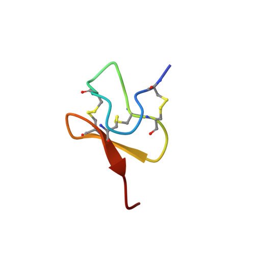
    Spider venom toxins, such as Protoxin-II (ProTx-II), have recently received much attention as selective Na v 1.7 channel blockers, with potential to be developed as leads for the treatment of chronic nocioceptive pain. ProTx-II is a 30-amino acid peptide with three disulfide bonds that has been reported to adopt a well-defined inhibitory cystine knot (ICK) scaffold structure. Potential drawbacks with such peptides include poor pharmacodynamics and potential scrambling of the disulfide bonds in vivo. In order to address these issues, in the present study we report the solid-phase synthesis of lanthionine-bridged analogues of ProTx-II, in which one of the three disulfide bridges is replaced with a thioether linkage, and evaluate the biological properties of these analogues. We have also investigated the folding and disulfide bridging patterns arising from different methods of oxidation of the linear peptide precursor. Finally, we report the X-ray crystal structure of ProTx-II to atomic resolution; to our knowledge this is the first crystal structure of an ICK spider venom peptide not bound to a substrate.


  • Organizational Affiliation
    • Department of Chemistry, University College London , 20 Gordon Street, London WC1H 0AJ, United Kingdom.

Macromolecule Content 

  • Total Structure Weight: 4.03 kDa 
  • Atom Count: 329 
  • Modeled Residue Count: 30 
  • Deposited Residue Count: 30 
  • Unique protein chains: 1

Macromolecules

Find similar proteins by:|  3D Structure
Entity ID: 1
MoleculeChains  Sequence LengthOrganismDetailsImage
Beta/omega-theraphotoxin-Tp2a30Thrixopelma pruriensMutation(s): 0 
UniProt
Find proteins for P83476 (Thrixopelma pruriens)
Explore P83476 
Go to UniProtKB:  P83476
Entity Groups
Sequence Clusters30% Identity50% Identity70% Identity90% Identity95% Identity100% Identity
UniProt GroupP83476
Sequence Annotations
Expand
Reference Sequence

Experimental Data & Validation

Experimental Data

  • Method: X-RAY DIFFRACTION
  • Resolution: 0.99 Å
  • R-Value Free:  0.140 (Depositor), 0.154 (DCC) 
  • R-Value Work:  0.126 (Depositor) 
  • R-Value Observed: 0.127 (Depositor) 
Space Group: H 3 2
Unit Cell:
Length ( Å )Angle ( ˚ )
a = 37.669α = 90
b = 37.669β = 90
c = 103.944γ = 120
Software Package:
Software NamePurpose
REFMACrefinement
SHELXLrefinement
SHARPphasing

Structure Validation

View Full Validation Report


Entry History 

& Funding Information

Deposition Data


Funding OrganizationLocationGrant Number
Wellcome TrustUnited Kingdom109073/Z/15/Z

Revision History  (Full details and data files)

  • Version 1.0: 2017-09-13
    Type: Initial release
  • Version 1.1: 2019-03-27
    Changes: Data collection, Database references
  • Version 1.2: 2024-10-16
    Changes: Data collection, Database references, Structure summary