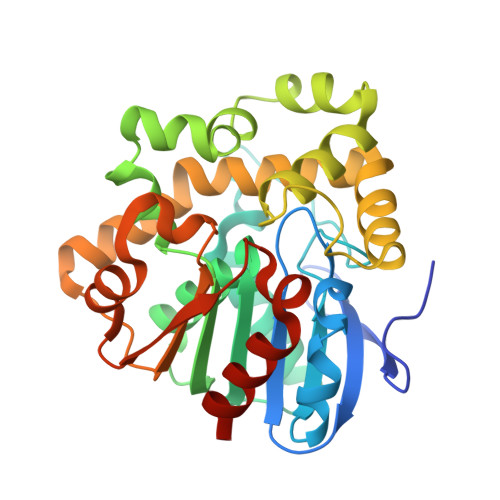
5NYV

Crystal structure determination from picosecond infrared laser ablated protein crystals by serial synchrotron crystallography


Experimental Data Snapshot

  • Method: X-RAY DIFFRACTION
  • Resolution: 1.60 Å
  • R-Value Free: 0.219 
  • R-Value Work: 0.193 
  • R-Value Observed: 0.193 

wwPDB Validation   3D Report Full Report


This is version 1.1 of the entry. See complete history


Literature

Protein crystals IR laser ablated from aqueous solution at high speed retain their diffractive properties: applications in high-speed serial crystallography.

Schulz, E.C.Kaub, J.Busse, F.Mehrabi, P.Muller-Werkmeister, H.M.Pai, E.F.Robertson, W.D.Miller, R.J.D.

(2017) J Appl Crystallogr 50: 1773-1781


Macromolecules
Find similar proteins by:  (by identity cutoff)  |  3D Structure
Entity ID: 1
MoleculeChains Sequence LengthOrganismDetailsImage
Fluoroacetate dehalogenase
A, B
306Rhodopseudomonas palustris ATCC 17001Mutation(s): 0 
Gene Names: RPA1163
EC: 3.8.1.3
UniProt
Find proteins for Q6NAM1 (Rhodopseudomonas palustris (strain ATCC BAA-98 / CGA009))
Explore Q6NAM1 
Go to UniProtKB:  Q6NAM1
Entity Groups  
Sequence Clusters30% Identity50% Identity70% Identity90% Identity95% Identity100% Identity
UniProt GroupQ6NAM1
Sequence Annotations
Expand
  • Reference Sequence
Experimental Data & Validation

Experimental Data

  • Method: X-RAY DIFFRACTION
  • Resolution: 1.60 Å
  • R-Value Free: 0.219 
  • R-Value Work: 0.193 
  • R-Value Observed: 0.193 
  • Space Group: P 1 21 1
Unit Cell:
Length ( Å )Angle ( ˚ )
a = 42.535α = 90
b = 80.889β = 102.72
c = 85.633γ = 90
Software Package:
Software NamePurpose
PHENIXrefinement
cctbx.xfeldata reduction
cctbx.primedata scaling
PHASERphasing

Structure Validation

View Full Validation Report


Entry History 

Deposition Data

Revision History  (Full details and data files)

  • Version 1.0: 2018-03-21
    Type: Initial release
  • Version 1.1: 2019-10-16
    Changes: Data collection