5NPU | pdb_00005npu

Inferred ancestral pyruvate decarboxylase


Experimental Data Snapshot

  • Method: X-RAY DIFFRACTION
  • Resolution: 3.50 Å
  • R-Value Free: 
    0.319 (Depositor), 0.324 (DCC) 
  • R-Value Work: 
    0.246 (Depositor), 0.248 (DCC) 
  • R-Value Observed: 
    0.250 (Depositor) 

Starting Model: experimental
View more details

wwPDB Validation 3D Report Full Report

Validation slider image for 5NPU

Ligand Structure Quality Assessment 


This is version 1.3 of the entry. See complete history

Literature

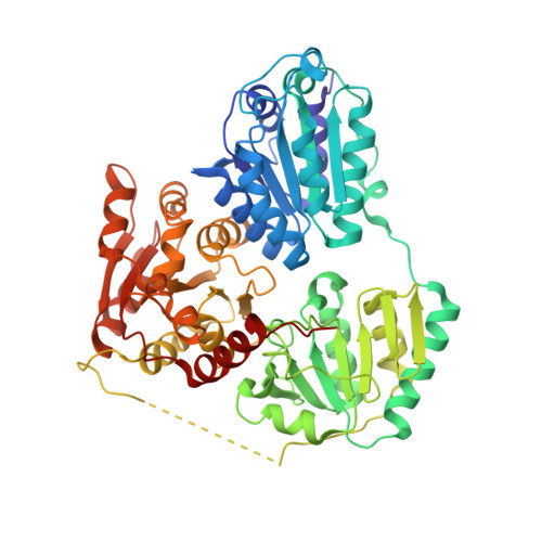
Crystal structure of an inferred ancestral bacterial pyruvate decarboxylase.

Buddrus, L.Andrews, E.S.V.Leak, D.J.Danson, M.J.Arcus, V.L.Crennell, S.J.

(2018) Acta Crystallogr F Struct Biol Commun 74: 179-186

  • DOI: https://doi.org/10.1107/S2053230X18002819
  • Primary Citation Related Structures: 
    5NPU

  • PubMed Abstract: 

    Pyruvate decarboxylase (PDC; EC 4.1.1.1) is a key enzyme in homofermentative metabolism where ethanol is the major product. PDCs are thiamine pyrophosphate- and Mg 2+ ion-dependent enzymes that catalyse the non-oxidative decarboxylation of pyruvate to acetaldehyde and carbon dioxide. As this enzyme class is rare in bacteria, current knowledge of bacterial PDCs is extremely limited. One approach to further the understanding of bacterial PDCs is to exploit the diversity provided by evolution. Ancestral sequence reconstruction (ASR) is a method of computational molecular evolution to infer extinct ancestral protein sequences, which can then be synthesized and experimentally characterized. Through ASR a novel PDC was generated, designated ANC27, that shares only 78% amino-acid sequence identity with its closest extant homologue (Komagataeibacter medellinensis PDC, GenBank accession No. WP_014105323.1), yet is fully functional. Crystals of this PDC diffracted to 3.5 Å resolution. The data were merged in space group P3 2 21, with unit-cell parameters a = b = 108.33, c = 322.65 Å, and contained two dimers (two tetramer halves) in the asymmetric unit. The structure was solved by molecular replacement using PDB entry 2wvg as a model, and the final R values were R work = 0.246 (0.3671 in the highest resolution bin) and R free = 0.319 (0.4482 in the highest resolution bin). Comparison with extant bacterial PDCs supports the previously observed correlation between decreased tetramer interface area (and number of interactions) and decreased thermostability.


  • Organizational Affiliation
    • School of Biochemistry, University of Bristol, University Walk, Bristol BS8 1TD, England.

Macromolecule Content 

  • Total Structure Weight: 244.71 kDa 
  • Atom Count: 16,656 
  • Modeled Residue Count: 2,182 
  • Deposited Residue Count: 2,252 
  • Unique protein chains: 1

Macromolecules

Find similar proteins by:|  3D Structure
Entity ID: 1
MoleculeChains  Sequence LengthOrganismDetailsImage
ANC27
A, B, C, D
563synthetic constructMutation(s): 0 

Small Molecules

Ligands 3 Unique
IDChains Name / Formula / InChI Key2D Diagram3D Interactions
TPP

Query on TPP


Download:Ideal Coordinates CCD File
F [auth A],
H [auth B],
J [auth C],
M [auth D]
THIAMINE DIPHOSPHATE
C12 H19 N4 O7 P2 S
AYEKOFBPNLCAJY-UHFFFAOYSA-O
PEG

Query on PEG


Download:Ideal Coordinates CCD File
K [auth C]DI(HYDROXYETHYL)ETHER
C4 H10 O3
MTHSVFCYNBDYFN-UHFFFAOYSA-N
MG

Query on MG


Download:Ideal Coordinates CCD File
E [auth A],
G [auth B],
I [auth C],
L [auth D]
MAGNESIUM ION
Mg
JLVVSXFLKOJNIY-UHFFFAOYSA-N

Experimental Data & Validation

Experimental Data

  • Method: X-RAY DIFFRACTION
  • Resolution: 3.50 Å
  • R-Value Free:  0.319 (Depositor), 0.324 (DCC) 
  • R-Value Work:  0.246 (Depositor), 0.248 (DCC) 
  • R-Value Observed: 0.250 (Depositor) 
Space Group: P 32 2 1
Unit Cell:
Length ( Å )Angle ( ˚ )
a = 108.33α = 90
b = 108.33β = 90
c = 322.651γ = 120
Software Package:
Software NamePurpose
PHENIXrefinement
iMOSFLMdata reduction
Aimlessdata scaling
BALBESphasing

Structure Validation

View Full Validation Report


Ligand Structure Quality Assessment 


Entry History 

Deposition Data

Revision History  (Full details and data files)

  • Version 1.0: 2018-03-07
    Type: Initial release
  • Version 1.1: 2018-04-18
    Changes: Data collection, Database references
  • Version 1.2: 2018-11-28
    Changes: Data collection, Source and taxonomy, Structure summary
  • Version 1.3: 2024-01-17
    Changes: Data collection, Database references, Derived calculations, Refinement description