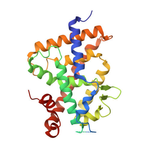
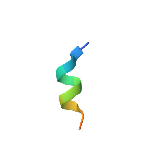
5NMB

Structure-activity relationship study of vitamin D analogs with oxolane group in their side chain


Experimental Data Snapshot

  • Method: X-RAY DIFFRACTION
  • Resolution: 2.50 Å
  • R-Value Free: 0.260 
  • R-Value Work: 0.197 
  • R-Value Observed: 0.200 

Starting Model: experimental
View more details

wwPDB Validation   3D Report Full Report


Ligand Structure Quality Assessment 

Created with Raphaël 2.3.0Worse 01 BetterLigand structure goodness of fit to experimental dataBest fitted 9CZClick on this verticalbar to view details

This is version 1.3 of the entry. See complete history


Literature

Structure-activity relationship study of vitamin D analogs with oxolane group in their side chain.

Belorusova, A.Y.Martinez, A.Gandara, Z.Gomez, G.Fall, Y.Rochel, N.

(2017) Eur J Med Chem 134: 86-96

  • DOI: https://doi.org/10.1016/j.ejmech.2017.03.081
  • Primary Citation of Related Structures:  
    5NKY, 5NMA, 5NMB

  • PubMed Abstract: 

    Synthetic analogs of 1α,25-dihydroxyvitamin D 3 (1,25(OH) 2 D 3 ) have been developed with the goal of improving the biological profile of the natural hormone for therapeutic applications. Derivatives of 1,25(OH) 2 D 3 with the oxolane moiety branched in the side chain at carbon C20, act as Vitamin D nuclear Receptor (VDR) superagonists being several orders of magnitude more active than the natural ligand. Here, we describe the synthesis and biological evaluation of three diastereoisomers of (1S, 3R)-Dihydroxy-(20S)-[(2″-hydroxy-2″-propyl)-tetrahydrofuryl]-22,23,24,25,26,27-hexanor-1α-hydroxyvitamin D3, with different stereochemistry at positions C2 and C5 of the oxolane ring branched at carbon C22 (1, C2RC5S; 2, C2SC5R; 3, C2SC5S). These compounds act as weak VDR agonist in transcriptional assays with compound 3 being the most active. X-ray crystallographic analysis of the VDR ligand-binding domain accommodating the three compounds indicates that the oxolane group branched at carbon C22 is not constrained as in case of compound with oxolane group branched at C20 leading to the loss of interactions of the triene group and increased flexibility of the C/D-rings and of the side chain.


  • Organizational Affiliation

    Department of Integrated Structural Biology, IGBMC (Institute of Genetics and of Molecular and Cellular Biology), 1 rue Laurent Fries, Illkirch, France; Centre National de la Recherche Scientifique (CNRS) UMR 7104, Illkirch, France; Institut National de la Santé et de la Recherche Médicale (INSERM) U964, Illkirch, France; Université de Strasbourg, Strasbourg, France.


Macromolecules
Find similar proteins by:  (by identity cutoff)  |  3D Structure
Entity ID: 1
MoleculeChains Sequence LengthOrganismDetailsImage
Vitamin D3 receptor AA [auth A2]302Danio rerioMutation(s): 0 
Gene Names: vdranr1i1avdr
UniProt
Find proteins for Q9PTN2 (Danio rerio)
Explore Q9PTN2 
Go to UniProtKB:  Q9PTN2
Entity Groups  
Sequence Clusters30% Identity50% Identity70% Identity90% Identity95% Identity100% Identity
UniProt GroupQ9PTN2
Sequence Annotations
Expand
  • Reference Sequence

Find similar proteins by:  Sequence   |   3D Structure  

Entity ID: 2
MoleculeChains Sequence LengthOrganismDetailsImage
Nuclear receptor coactivator 1B [auth B2]15Homo sapiensMutation(s): 0 
EC: 2.3.1.48
UniProt & NIH Common Fund Data Resources
Find proteins for Q15788 (Homo sapiens)
Explore Q15788 
Go to UniProtKB:  Q15788
PHAROS:  Q15788
GTEx:  ENSG00000084676 
Entity Groups  
Sequence Clusters30% Identity50% Identity70% Identity90% Identity95% Identity100% Identity
UniProt GroupQ15788
Sequence Annotations
Expand
  • Reference Sequence
Small Molecules
Ligands 1 Unique
IDChains Name / Formula / InChI Key2D Diagram3D Interactions
9CZ
Query on 9CZ

Download Ideal Coordinates CCD File 
C [auth A2](1~{R},3~{S},5~{Z})-5-[(2~{E})-2-[(1~{R},3~{a}~{S},7~{a}~{R})-7~{a}-methyl-1-[(1~{S})-1-[(2~{S},5~{R})-5-(2-oxidanylpropan-2-yl)oxolan-2-yl]ethyl]-2,3,3~{a},5,6,7-hexahydro-1~{H}-inden-4-ylidene]ethylidene]-4-methylidene-cyclohexane-1,3-diol
C28 H44 O4
DDIWZQGTNVZJGO-XOJGNBOJSA-N
Experimental Data & Validation

Experimental Data

  • Method: X-RAY DIFFRACTION
  • Resolution: 2.50 Å
  • R-Value Free: 0.260 
  • R-Value Work: 0.197 
  • R-Value Observed: 0.200 
  • Space Group: P 65 2 2
Unit Cell:
Length ( Å )Angle ( ˚ )
a = 65.893α = 90
b = 65.893β = 90
c = 262.598γ = 120
Software Package:
Software NamePurpose
PHENIXrefinement
HKL-2000data reduction
HKL-2000data scaling
PHENIXphasing

Structure Validation

View Full Validation Report


Ligand Structure Quality Assessment 

Created with Raphaël 2.3.0Worse 01 BetterLigand structure goodness of fit to experimental dataBest fitted 9CZClick on this verticalbar to view details

Entry History & Funding Information

Deposition Data


Funding OrganizationLocationGrant Number
French National Research AgencyFranceANR-13-BSV8-0024-01
FRMFranceFRM: FDT20140930978

Revision History  (Full details and data files)

  • Version 1.0: 2017-05-24
    Type: Initial release
  • Version 1.1: 2017-06-07
    Changes: Other
  • Version 1.2: 2017-09-06
    Changes: Author supporting evidence
  • Version 1.3: 2024-01-17
    Changes: Advisory, Data collection, Database references, Refinement description