5NLB | pdb_00005nlb

Crystal structure of human CUL3 N-terminal domain bound to KEAP1 BTB and 3-box


Experimental Data Snapshot

  • Method: X-RAY DIFFRACTION
  • Resolution: 3.45 Å
  • R-Value Free: 
    0.288 (Depositor), 0.297 (DCC) 
  • R-Value Work: 
    0.238 (Depositor), 0.242 (DCC) 
  • R-Value Observed: 
    0.241 (Depositor) 

Starting Model: experimental
View more details

wwPDB Validation 3D Report Full Report

Validation slider image for 5NLB

This is version 1.4 of the entry. See complete history

Literature

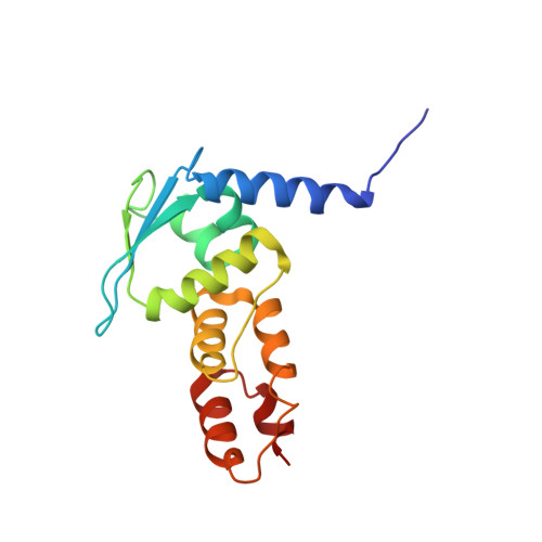
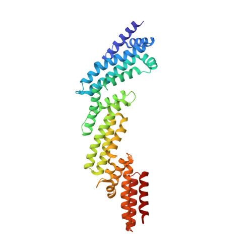
Structural and biochemical characterization establishes a detailed understanding of KEAP1-CUL3 complex assembly.

Adamson, R.J.Payne, N.C.Bartual, S.G.Mazitschek, R.Bullock, A.N.

(2023) Free Radic Biol Med 204: 215-225

  • DOI: https://doi.org/10.1016/j.freeradbiomed.2023.04.021
  • Primary Citation Related Structures: 
    5NLB

  • PubMed Abstract: 

    KEAP1 promotes the ubiquitin-dependent degradation of NRF2 by assembling into a CUL3-dependent ubiquitin ligase complex. Oxidative and electrophilic stress inhibit KEAP1 allowing NRF2 to accumulate for the transactivation of stress response genes. To date there are no structures of the KEAP1-CUL3 interaction nor binding data to show the contributions of different domains to their binding affinity. We determined a crystal structure of the BTB and 3-box domains of human KEAP1 in complex with the CUL3 N-terminal domain that showed a heterotetrameric assembly with 2:2 stoichiometry. To support the structural data, we developed a versatile TR-FRET-based assay system to profile the binding of BTB-domain-containing proteins to CUL3 and determine the contribution of distinct protein features, revealing the importance of the CUL3 N-terminal extension for high affinity binding. We further provide direct evidence that the investigational drug CDDO does not disrupt the KEAP1-CUL3 interaction, even at high concentrations, but reduces the affinity of KEAP1-CUL3 binding. The TR-FRET-based assay system offers a generalizable platform for profiling this protein class and may form a suitable screening platform for ligands that disrupt these interactions by targeting the BTB or 3-box domains to block E3 ligase function.


  • Organizational Affiliation
    • Centre for Medicines Discovery, Nuffield Department of Medicine, University of Oxford, Oxford, OX3 7FZ, UK.

Macromolecule Content 

  • Total Structure Weight: 58.9 kDa 
  • Atom Count: 3,882 
  • Modeled Residue Count: 502 
  • Deposited Residue Count: 510 
  • Unique protein chains: 2

Macromolecules

Find similar proteins by:|  3D Structure
Entity ID: 1
MoleculeChains  Sequence LengthOrganismDetailsImage
Kelch-like ECH-associated protein 1154Homo sapiensMutation(s): 0 
Gene Names: KEAP1INRF2KIAA0132KLHL19
UniProt & NIH Common Fund Data Resources
Find proteins for Q14145 (Homo sapiens)
Explore Q14145 
Go to UniProtKB:  Q14145
PHAROS:  Q14145
GTEx:  ENSG00000079999 
Entity Groups
Sequence Clusters30% Identity50% Identity70% Identity90% Identity95% Identity100% Identity
UniProt GroupQ14145
Sequence Annotations
Expand
Reference Sequence
Find similar proteins by:|  3D Structure
Entity ID: 2
MoleculeChains  Sequence LengthOrganismDetailsImage
Cullin-3356Homo sapiensMutation(s): 2 
Gene Names: CUL3KIAA0617
UniProt & NIH Common Fund Data Resources
Find proteins for Q13618 (Homo sapiens)
Explore Q13618 
Go to UniProtKB:  Q13618
PHAROS:  Q13618
GTEx:  ENSG00000036257 
Entity Groups
Sequence Clusters30% Identity50% Identity70% Identity90% Identity95% Identity100% Identity
UniProt GroupQ13618
Sequence Annotations
Expand
Reference Sequence

Experimental Data & Validation

Experimental Data

  • Method: X-RAY DIFFRACTION
  • Resolution: 3.45 Å
  • R-Value Free:  0.288 (Depositor), 0.297 (DCC) 
  • R-Value Work:  0.238 (Depositor), 0.242 (DCC) 
  • R-Value Observed: 0.241 (Depositor) 
Space Group: C 2 2 21
Unit Cell:
Length ( Å )Angle ( ˚ )
a = 41α = 90
b = 233.38β = 90
c = 164.71γ = 90
Software Package:
Software NamePurpose
PHENIXrefinement
xia2data reduction
xia2data scaling
PHENIXphasing

Structure Validation

View Full Validation Report


Entry History 

Revision History  (Full details and data files)

  • Version 1.0: 2017-04-19
    Type: Initial release
  • Version 1.1: 2017-08-02
    Changes: Database references, Structure summary
  • Version 1.2: 2019-10-16
    Changes: Data collection
  • Version 1.3: 2023-11-08
    Changes: Data collection, Database references
  • Version 1.4: 2024-01-17
    Changes: Refinement description