5NJB

E. coli Microcin-processing metalloprotease TldD/E with actinonin bound


Experimental Data Snapshot

  • Method: X-RAY DIFFRACTION
  • Resolution: 1.50 Å
  • R-Value Free: 0.169 
  • R-Value Work: 0.130 
  • R-Value Observed: 0.132 

Starting Model: experimental
View more details

wwPDB Validation   3D Report Full Report


Ligand Structure Quality Assessment 


This is version 1.2 of the entry. See complete history


Literature

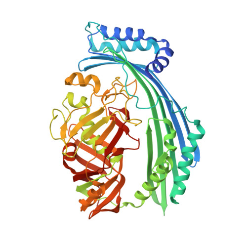
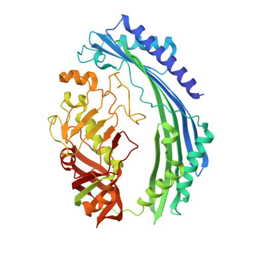
The Origins of Specificity in the Microcin-Processing Protease TldD/E.

Ghilarov, D.Serebryakova, M.Stevenson, C.E.M.Hearnshaw, S.J.Volkov, D.S.Maxwell, A.Lawson, D.M.Severinov, K.

(2017) Structure 25: 1549-1561.e5

  • DOI: https://doi.org/10.1016/j.str.2017.08.006
  • Primary Citation of Related Structures:  
    5NJ5, 5NJ9, 5NJA, 5NJB, 5NJC, 5NJF

  • PubMed Abstract: 

    TldD and TldE proteins are involved in the biosynthesis of microcin B17 (MccB17), an Escherichia coli thiazole/oxazole-modified peptide toxin targeting DNA gyrase. Using a combination of biochemical and crystallographic methods we show that E. coli TldD and TldE interact to form a heterodimeric metalloprotease. TldD/E cleaves the N-terminal leader sequence from the modified MccB17 precursor peptide, to yield mature antibiotic, while it has no effect on the unmodified peptide. Both proteins are essential for the activity; however, only the TldD subunit forms a novel metal-containing active site within the hollow core of the heterodimer. Peptide substrates are bound in a sequence-independent manner through β sheet interactions with TldD and are likely cleaved via a thermolysin-type mechanism. We suggest that TldD/E acts as a "molecular pencil sharpener": unfolded polypeptides are fed through a narrow channel into the active site and processively truncated through the cleavage of short peptides from the N-terminal end.


  • Organizational Affiliation

    Centre for Data-Intensive Biomedicine and Biotechnology, Skolkovo Institute of Science and Technology, 143026 Moscow, Russia; Institute of Gene Biology of the Russian Academy of Sciences, 119334 Moscow, Russia; Department of Biological Chemistry, John Innes Centre, Norwich Research Park, Norwich NR4 7UH, UK. Electronic address: dmitry.gilyarov@uj.edu.pl.


Macromolecules
Find similar proteins by:  (by identity cutoff)  |  3D Structure
Entity ID: 1
MoleculeChains Sequence LengthOrganismDetailsImage
Metalloprotease TldD
A, C
495Escherichia coli K-12Mutation(s): 1 
Gene Names: tldDyhdOb3244JW3213
EC: 3.4
UniProt
Find proteins for P0AGG8 (Escherichia coli (strain K12))
Explore P0AGG8 
Go to UniProtKB:  P0AGG8
Entity Groups  
Sequence Clusters30% Identity50% Identity70% Identity90% Identity95% Identity100% Identity
UniProt GroupP0AGG8
Sequence Annotations
Expand
  • Reference Sequence
Find similar proteins by:  (by identity cutoff)  |  3D Structure
Entity ID: 2
MoleculeChains Sequence LengthOrganismDetailsImage
Metalloprotease PmbA
B, D
450Escherichia coli K-12Mutation(s): 0 
Gene Names: pmbAtldEb4235JW4194
EC: 3.4
UniProt
Find proteins for P0AFK0 (Escherichia coli (strain K12))
Explore P0AFK0 
Go to UniProtKB:  P0AFK0
Entity Groups  
Sequence Clusters30% Identity50% Identity70% Identity90% Identity95% Identity100% Identity
UniProt GroupP0AFK0
Sequence Annotations
Expand
  • Reference Sequence
Small Molecules
Ligands 4 Unique
IDChains Name / Formula / InChI Key2D Diagram3D Interactions
BB2
Query on BB2

Download Ideal Coordinates CCD File 
F [auth A],
M [auth C]
ACTINONIN
C19 H35 N3 O5
XJLATMLVMSFZBN-VYDXJSESSA-N
MES
Query on MES

Download Ideal Coordinates CCD File 
G [auth A],
N [auth C]
2-(N-MORPHOLINO)-ETHANESULFONIC ACID
C6 H13 N O4 S
SXGZJKUKBWWHRA-UHFFFAOYSA-N
ZN
Query on ZN

Download Ideal Coordinates CCD File 
E [auth A],
L [auth C]
ZINC ION
Zn
PTFCDOFLOPIGGS-UHFFFAOYSA-N
EDO
Query on EDO

Download Ideal Coordinates CCD File 
H [auth A]
I [auth A]
J [auth B]
K [auth B]
O [auth C]
H [auth A],
I [auth A],
J [auth B],
K [auth B],
O [auth C],
P [auth D],
Q [auth D],
R [auth D],
S [auth D]
1,2-ETHANEDIOL
C2 H6 O2
LYCAIKOWRPUZTN-UHFFFAOYSA-N
Experimental Data & Validation

Experimental Data

  • Method: X-RAY DIFFRACTION
  • Resolution: 1.50 Å
  • R-Value Free: 0.169 
  • R-Value Work: 0.130 
  • R-Value Observed: 0.132 
  • Space Group: P 1 21 1
Unit Cell:
Length ( Å )Angle ( ˚ )
a = 64.57α = 90
b = 172.88β = 90.02
c = 82.63γ = 90
Software Package:
Software NamePurpose
Aimlessdata scaling
REFMACrefinement
PDB_EXTRACTdata extraction
XDSdata reduction
REFMACphasing

Structure Validation

View Full Validation Report


Ligand Structure Quality Assessment 


Entry History & Funding Information

Deposition Data


Funding OrganizationLocationGrant Number
Dynasty foundationRussian FederationPersonal fellowship
Russian Ministry of ScienceRussian FederationResearch and Scientific-Pedagogical Personnel of Innovative Russia in 2009-2013 grant 8591
Moscow State UniversityRussian FederationDevelopment Program PNG 5.13
European UnionPolandMarie Sklodowska-Curie grant agreement No. 665778
Biotechnology and Biological Sciences Research CouncilUnited KingdomBB/J016853/1
Biotechnology and Biological Sciences Research CouncilUnited KingdomBB/J004561/1 (MET)
John Innes FoundationUnited Kingdom--

Revision History  (Full details and data files)

  • Version 1.0: 2017-10-04
    Type: Initial release
  • Version 1.1: 2017-10-18
    Changes: Database references
  • Version 1.2: 2024-01-17
    Changes: Data collection, Database references, Derived calculations, Refinement description