5NGZ | pdb_00005ngz

Ube2T in complex with fragment EM04


Experimental Data Snapshot

  • Method: X-RAY DIFFRACTION
  • Resolution: 2.40 Å
  • R-Value Free: 
    0.247 (Depositor), 0.252 (DCC) 
  • R-Value Work: 
    0.210 (Depositor), 0.215 (DCC) 
  • R-Value Observed: 
    0.212 (Depositor) 

Starting Model: experimental
View more details

wwPDB Validation 3D Report Full Report

Validation slider image for 5NGZ

Ligand Structure Quality Assessment 


This is version 1.2 of the entry. See complete history

Literature

Allosteric Targeting of the Fanconi Anemia Ubiquitin-Conjugating Enzyme Ube2T by Fragment Screening.

Morreale, F.E.Bortoluzzi, A.Chaugule, V.K.Arkinson, C.Walden, H.Ciulli, A.

(2017) J Med Chem 60: 4093-4098

  • DOI: https://doi.org/10.1021/acs.jmedchem.7b00147
  • Primary Citation Related Structures: 
    5NGZ

  • PubMed Abstract: 

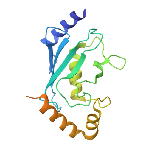
    Ube2T is the E2 ubiquitin-conjugating enzyme of the Fanconi anemia DNA repair pathway and it is overexpressed in several cancers, representing an attractive target for the development of inhibitors. Despite the extensive efforts in targeting the ubiquitin system, very few E2 binders have currently been discovered. Herein we report the identification of a new allosteric pocket on Ube2T through a fragment screening using biophysical methods. Several fragments binding to this site inhibit ubiquitin conjugation in vitro.


  • Organizational Affiliation
    • MRC Protein Phosphorylation and Ubiquitylation Unit, School of Life Sciences, University of Dundee , Dundee DD1 5EH, United Kingdom.

Macromolecule Content 

  • Total Structure Weight: 22.72 kDa 
  • Atom Count: 1,276 
  • Modeled Residue Count: 152 
  • Deposited Residue Count: 197 
  • Unique protein chains: 1

Macromolecules

Find similar proteins by:|  3D Structure
Entity ID: 1
MoleculeChains  Sequence LengthOrganismDetailsImage
Ubiquitin-conjugating enzyme E2 T197Homo sapiensMutation(s): 0 
Gene Names: UBE2THSPC150PIG50
EC: 2.3.2.23
UniProt & NIH Common Fund Data Resources
Find proteins for Q9NPD8 (Homo sapiens)
Explore Q9NPD8 
Go to UniProtKB:  Q9NPD8
PHAROS:  Q9NPD8
GTEx:  ENSG00000077152 
Entity Groups
Sequence Clusters30% Identity50% Identity70% Identity90% Identity95% Identity100% Identity
UniProt GroupQ9NPD8
Sequence Annotations
Expand
Reference Sequence

Small Molecules

Ligands 1 Unique
IDChains Name / Formula / InChI Key2D Diagram3D Interactions
2BG

Query on 2BG


Download:Ideal Coordinates CCD File
B [auth A]1-(1,3-benzothiazol-2-yl)methanamine
C8 H8 N2 S
VLBUERZRFSORRZ-UHFFFAOYSA-N
Binding Affinity Annotations 
IDSourceBinding Affinity
2BG BindingDB:  5NGZ Kd: 1.30e+6 (nM) from 1 assay(s)

Experimental Data & Validation

Experimental Data

  • Method: X-RAY DIFFRACTION
  • Resolution: 2.40 Å
  • R-Value Free:  0.247 (Depositor), 0.252 (DCC) 
  • R-Value Work:  0.210 (Depositor), 0.215 (DCC) 
  • R-Value Observed: 0.212 (Depositor) 
Space Group: P 43 21 2
Unit Cell:
Length ( Å )Angle ( ˚ )
a = 55.817α = 90
b = 55.817β = 90
c = 185.278γ = 90
Software Package:
Software NamePurpose
REFMACrefinement
XDSdata reduction
Aimlessdata scaling
REFMACphasing

Structure Validation

View Full Validation Report


Ligand Structure Quality Assessment 


Entry History 

Deposition Data

Revision History  (Full details and data files)

  • Version 1.0: 2017-05-03
    Type: Initial release
  • Version 1.1: 2017-05-24
    Changes: Database references
  • Version 1.2: 2024-01-17
    Changes: Data collection, Database references, Refinement description