5NFF | pdb_00005nff

Crystal structure of GP1 receptor binding domain from Morogoro virus


Experimental Data Snapshot

  • Method: X-RAY DIFFRACTION
  • Resolution: 2.62 Å
  • R-Value Free: 
    0.207 (Depositor), 0.217 (DCC) 
  • R-Value Work: 
    0.174 (Depositor), 0.186 (DCC) 
  • R-Value Observed: 
    0.176 (Depositor) 

Starting Model: experimental
View more details

wwPDB Validation 3D Report Full Report

Validation slider image for 5NFF

Ligand Structure Quality Assessment 


This is version 2.2 of the entry. See complete history

Literature

Mapping of the Lassa virus LAMP1 binding site reveals unique determinants not shared by other old world arenaviruses.

Israeli, H.Cohen-Dvashi, H.Shulman, A.Shimon, A.Diskin, R.

(2017) PLoS Pathog 13: e1006337-e1006337

  • DOI: https://doi.org/10.1371/journal.ppat.1006337
  • Primary Citation Related Structures: 
    5NFF

  • PubMed Abstract: 

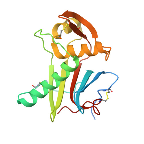
    Cell entry of many enveloped viruses occurs by engagement with cellular receptors, followed by internalization into endocytic compartments and pH-induced membrane fusion. A previously unnoticed step of receptor switching was found to be critical during cell entry of two devastating human pathogens: Ebola and Lassa viruses. Our recent studies revealed the functional role of receptor switching to LAMP1 for triggering membrane fusion by Lassa virus and showed the involvement of conserved histidines in this switching, suggesting that other viruses from this family may also switch to LAMP1. However, when we investigated viruses that are genetically close to Lassa virus, we discovered that they cannot bind LAMP1. A crystal structure of the receptor-binding module from Morogoro virus revealed structural differences that allowed mapping of the LAMP1 binding site to a unique set of Lassa residues not shared by other viruses in its family, illustrating a key difference in the cell-entry mechanism of Lassa virus that may contribute to its pathogenicity.


  • Organizational Affiliation
    • Department of Structural Biology, Weizmann Institute of Science, Rehovot, Israel.

Macromolecule Content 

  • Total Structure Weight: 317.55 kDa 
  • Atom Count: 20,391 
  • Modeled Residue Count: 2,496 
  • Deposited Residue Count: 2,736 
  • Unique protein chains: 1

Macromolecules

Find similar proteins by:|  3D Structure
Entity ID: 1
MoleculeChains  Sequence LengthOrganismDetailsImage
Glycoprotein171Morogoro mammarenavirusMutation(s): 0 
Gene Names: GPC
UniProt
Find proteins for C6ZK00 (Morogoro mammarenavirus)
Explore C6ZK00 
Go to UniProtKB:  C6ZK00
Entity Groups
Sequence Clusters30% Identity50% Identity70% Identity90% Identity95% Identity100% Identity
UniProt GroupC6ZK00
Glycosylation
Glycosylation Sites: 1
Sequence Annotations
Expand
Reference Sequence

Oligosaccharides

Help  
Entity ID: 2
MoleculeChains Length2D Diagram GlycosylationD Interactions
2-acetamido-2-deoxy-beta-D-glucopyranose-(1-4)-2-acetamido-2-deoxy-beta-D-glucopyranose
Q, R
2N-Glycosylation
Glycosylation Resources
GlyTouCan: G42666HT
GlyCosmos: G42666HT
GlyGen: G42666HT
Entity ID: 3
MoleculeChains Length2D Diagram GlycosylationD Interactions
alpha-D-mannopyranose-(1-6)-alpha-D-mannopyranose-(1-4)-2-acetamido-2-deoxy-beta-D-glucopyranose-(1-4)-2-acetamido-2-deoxy-beta-D-glucopyranose
S
4N-Glycosylation
Glycosylation Resources
GlyTouCan: G04854NQ
GlyCosmos: G04854NQ
GlyGen: G04854NQ
Entity ID: 4
MoleculeChains Length2D Diagram GlycosylationD Interactions
alpha-D-mannopyranose-(1-2)-alpha-D-mannopyranose-(1-3)-alpha-D-mannopyranose-(1-4)-2-acetamido-2-deoxy-beta-D-glucopyranose-(1-4)-2-acetamido-2-deoxy-beta-D-glucopyranose
T
5N-Glycosylation
Glycosylation Resources
GlyTouCan: G75436UI
GlyCosmos: G75436UI
GlyGen: G75436UI

Small Molecules

Ligands 2 Unique
IDChains Name / Formula / InChI Key2D Diagram3D Interactions
NAG

Query on NAG


Download:Ideal Coordinates CCD File
CA [auth D]
DA [auth E]
EA [auth E]
HA [auth G]
IA [auth G]
CA [auth D],
DA [auth E],
EA [auth E],
HA [auth G],
IA [auth G],
JA [auth H],
KA [auth I],
MA [auth J],
NA [auth K],
OA [auth L],
U [auth B],
V [auth B],
X [auth A],
Y [auth A],
Z [auth C]
2-acetamido-2-deoxy-beta-D-glucopyranose
C8 H15 N O6
OVRNDRQMDRJTHS-FMDGEEDCSA-N
CIT

Query on CIT


Download:Ideal Coordinates CCD File
AA [auth C]
BA [auth C]
FA [auth E]
GA [auth F]
LA [auth I]
AA [auth C],
BA [auth C],
FA [auth E],
GA [auth F],
LA [auth I],
W [auth B]
CITRIC ACID
C6 H8 O7
KRKNYBCHXYNGOX-UHFFFAOYSA-N

Experimental Data & Validation

Experimental Data

  • Method: X-RAY DIFFRACTION
  • Resolution: 2.62 Å
  • R-Value Free:  0.207 (Depositor), 0.217 (DCC) 
  • R-Value Work:  0.174 (Depositor), 0.186 (DCC) 
  • R-Value Observed: 0.176 (Depositor) 
Space Group: P 32
Unit Cell:
Length ( Å )Angle ( ˚ )
a = 127.769α = 90
b = 127.769β = 90
c = 251.704γ = 120
Software Package:
Software NamePurpose
PHENIXrefinement
PDB_EXTRACTdata extraction
XDSdata reduction
XSCALEdata scaling
PHASERphasing

Structure Validation

View Full Validation Report


Ligand Structure Quality Assessment 


Entry History 

& Funding Information

Deposition Data


Funding OrganizationLocationGrant Number
ISF - ICOREIsrael1775/12

Revision History  (Full details and data files)

  • Version 1.0: 2017-04-19
    Type: Initial release
  • Version 1.1: 2017-05-10
    Changes: Database references
  • Version 1.2: 2019-08-14
    Changes: Data collection
  • Version 2.0: 2020-07-29
    Type: Remediation
    Reason: Carbohydrate remediation
    Changes: Advisory, Atomic model, Data collection, Derived calculations, Structure summary
  • Version 2.1: 2024-01-17
    Changes: Data collection, Database references, Derived calculations, Refinement description, Structure summary
  • Version 2.2: 2024-11-20
    Changes: Structure summary