5NE9 | pdb_00005ne9

Crystal structure of H60A H307A mutant of Thermotoga maritima TmPEP1050 aminopeptidase


Experimental Data Snapshot

  • Method: X-RAY DIFFRACTION
  • Resolution: 2.37 Å
  • R-Value Free: 
    0.234 (Depositor), 0.232 (DCC) 
  • R-Value Work: 
    0.206 (Depositor), 0.208 (DCC) 
  • R-Value Observed: 
    0.209 (Depositor) 

Starting Model: experimental
View more details

wwPDB Validation 3D Report Full Report

Validation slider image for 5NE9

This is version 1.3 of the entry. See complete history

Literature

X-Ray Crystallography to Study the Oligomeric State Transition of the Thermotoga maritima M42 Aminopeptidase TmPep1050.

Dutoit, R.Brandt, N.Van Elder, D.Droogmans, L.

(2020) J Vis Exp 

  • DOI: https://doi.org/10.3791/61288
  • Primary Citation Related Structures: 
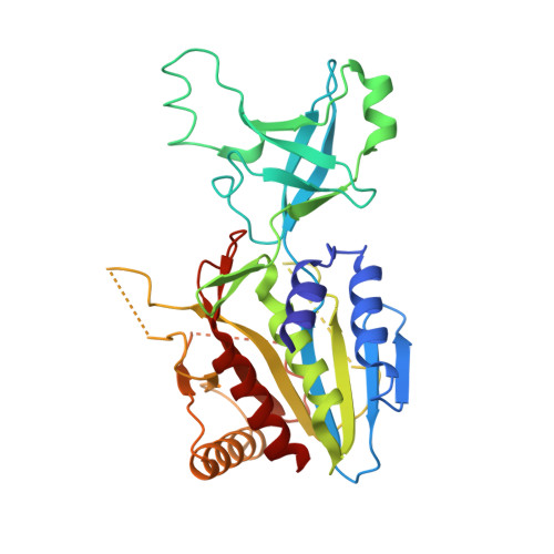
    5NE9

  • PubMed Abstract: 

    The M42 aminopeptidases form functionally active complexes made of 12 subunits. Their assembly process appears to be regulated by their metal ion cofactors triggering a dimer-dodecamer transition. Upon metal ion binding, several structural modifications occur in the active site and at the interaction interface, shaping dimers to promote the self-assembly. To observe such modifications, stable oligomers must be isolated prior to structural study. Reported here is a method that allows the purification of stable dodecamers and dimers of TmPep1050, an M42 aminopeptidase of T. maritima, and their structure determination by X-ray crystallography. Dimers were prepared from dodecamers by removing metal ions with a chelating agent. Without their cofactor, dodecamers became less stable and were fully dissociated upon heating. The oligomeric structures were solved by the straightforward molecular replacement approach. To illustrate the methodology, the structure of a TmPep1050 variant, totally impaired in metal ion binding, is presented showing no further breakdown of dimers to monomers.


  • Organizational Affiliation
    • Laboratory of Microbiology, Department of Molecular Biology, Université Libre de Bruxelles; Labiris Institut de Recherche; Raphael.Dutoit@ulb.ac.be.

Macromolecule Content 

  • Total Structure Weight: 71.95 kDa 
  • Atom Count: 4,655 
  • Modeled Residue Count: 616 
  • Deposited Residue Count: 662 
  • Unique protein chains: 1

Macromolecules

Find similar proteins by:|  3D Structure
Entity ID: 1
MoleculeChains  Sequence LengthOrganismDetailsImage
AMINOPEPTIDASE
A, B
331Thermotoga maritima MSB8Mutation(s): 2 
Gene Names: TM_1050Tmari_1054
EC: 3.4.11.1 (PDB Primary Data), 3.2.1.4 (UniProt)
UniProt
Find proteins for Q9X0E0 (Thermotoga maritima (strain ATCC 43589 / DSM 3109 / JCM 10099 / NBRC 100826 / MSB8))
Explore Q9X0E0 
Go to UniProtKB:  Q9X0E0
Entity Groups
Sequence Clusters30% Identity50% Identity70% Identity90% Identity95% Identity100% Identity
UniProt GroupQ9X0E0
Sequence Annotations
Expand
Reference Sequence

Experimental Data & Validation

Experimental Data

  • Method: X-RAY DIFFRACTION
  • Resolution: 2.37 Å
  • R-Value Free:  0.234 (Depositor), 0.232 (DCC) 
  • R-Value Work:  0.206 (Depositor), 0.208 (DCC) 
  • R-Value Observed: 0.209 (Depositor) 
Space Group: P 1 21 1
Unit Cell:
Length ( Å )Angle ( ˚ )
a = 43.24α = 90
b = 137.79β = 110.69
c = 61.11γ = 90
Software Package:
Software NamePurpose
XSCALEdata scaling
PHASERphasing
PHENIXrefinement
PDB_EXTRACTdata extraction
XDSdata reduction

Structure Validation

View Full Validation Report


Entry History 

& Funding Information

Deposition Data

  • Released Date: 2018-05-16 
  • Deposition Author(s): Dutoit, R.

Funding OrganizationLocationGrant Number
BAG-BelgiumBelgium20151139

Revision History  (Full details and data files)

  • Version 1.0: 2018-05-16
    Type: Initial release
  • Version 1.1: 2019-12-25
    Changes: Data collection
  • Version 1.2: 2021-02-24
    Changes: Database references
  • Version 1.3: 2024-01-17
    Changes: Data collection, Database references, Refinement description