5NAN | pdb_00005nan

Crystal Structure of human IL-17AF in complex with human IL-17RA


Experimental Data Snapshot

  • Method: X-RAY DIFFRACTION
  • Resolution: 3.30 Å
  • R-Value Free: 
    0.243 (Depositor), 0.270 (DCC) 
  • R-Value Work: 
    0.186 (Depositor), 0.218 (DCC) 
  • R-Value Observed: 
    0.189 (Depositor) 

Starting Model: experimental
View more details

wwPDB Validation 3D Report Full Report

Validation slider image for 5NAN

Ligand Structure Quality Assessment 


This is version 2.2 of the entry. See complete history

Literature

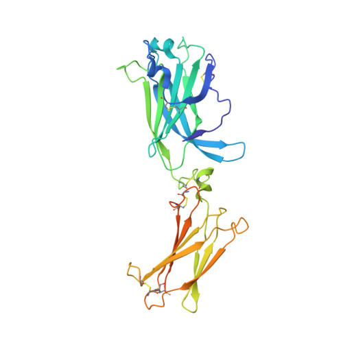
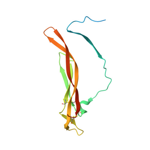
The human IL-17A/F heterodimer: a two-faced cytokine with unique receptor recognition properties.

Goepfert, A.Lehmann, S.Wirth, E.Rondeau, J.M.

(2017) Sci Rep 7: 8906-8906

  • DOI: https://doi.org/10.1038/s41598-017-08360-9
  • Primary Citation Related Structures: 
    5N92, 5NAN

  • PubMed Abstract: 

    IL-17A and IL-17F are prominent members of the IL-17 family of cytokines that regulates both innate and adaptive immunity. IL-17A has been implicated in chronic inflammatory and autoimmune diseases, and anti-IL-17A antibodies have shown remarkable clinical efficacy in psoriasis and psoriatic arthritis patients. IL-17A and IL-17F are homodimeric cytokines that can also form the IL-17A/F heterodimer whose precise role in health and disease remains elusive. All three cytokines signal through the assembly of a ternary complex with the IL-17RA and IL-17RC receptors. Here we report the X-ray analysis of the human IL-17A/F heterodimer that reveals a two-faced cytokine closely mimicking IL-17A as well as IL-17F. We also present the crystal structure of its complex with the IL-17RA receptor. Unexpectedly in view of the much higher affinity of this receptor toward IL-17A, we find that IL-17RA is bound to the "F-face" of the heterodimer in the crystal. Using site-directed mutagenesis, we then demonstrate that IL-17RA can also bind to the "A-face" of IL-17A/F with similar affinity. Further, we show that IL-17RC does not discriminate between the two faces of the cytokine heterodimer either, thus enabling the formation of two topologically-distinct heterotrimeric complexes with potentially different signaling properties.


  • Organizational Affiliation
    • Novartis Institutes for BioMedical Research, Novartis Pharma AG, CH-4002, Basel, Switzerland.

Macromolecule Content 

  • Total Structure Weight: 135.78 kDa 
  • Atom Count: 7,923 
  • Modeled Residue Count: 972 
  • Deposited Residue Count: 1,164 
  • Unique protein chains: 3

Macromolecules

Find similar proteins by:|  3D Structure
Entity ID: 1
MoleculeChains  Sequence LengthOrganismDetailsImage
Interleukin-17A
A, D
132Homo sapiensMutation(s): 0 
Gene Names: IL17ACTLA8IL17
UniProt & NIH Common Fund Data Resources
Find proteins for Q16552 (Homo sapiens)
Explore Q16552 
Go to UniProtKB:  Q16552
PHAROS:  Q16552
GTEx:  ENSG00000112115 
Entity Groups
Sequence Clusters30% Identity50% Identity70% Identity90% Identity95% Identity100% Identity
UniProt GroupQ16552
Sequence Annotations
Expand
Reference Sequence
Find similar proteins by:|  3D Structure
Entity ID: 2
MoleculeChains  Sequence LengthOrganismDetailsImage
Interleukin-17 receptor A
B, C
311Homo sapiensMutation(s): 0 
Gene Names: IL17RAIL17R
UniProt & NIH Common Fund Data Resources
Find proteins for Q96F46 (Homo sapiens)
Explore Q96F46 
Go to UniProtKB:  Q96F46
PHAROS:  Q96F46
GTEx:  ENSG00000177663 
Entity Groups
Sequence Clusters30% Identity50% Identity70% Identity90% Identity95% Identity100% Identity
UniProt GroupQ96F46
Glycosylation
Glycosylation Sites: 3Go to GlyGen: Q96F46-1
Sequence Annotations
Expand
Reference Sequence
Find similar proteins by:|  3D Structure
Entity ID: 3
MoleculeChains  Sequence LengthOrganismDetailsImage
Interleukin-17F
E, F
139Homo sapiensMutation(s): 0 
Gene Names: IL17F
UniProt & NIH Common Fund Data Resources
Find proteins for Q96PD4 (Homo sapiens)
Explore Q96PD4 
Go to UniProtKB:  Q96PD4
PHAROS:  Q96PD4
GTEx:  ENSG00000112116 
Entity Groups
Sequence Clusters30% Identity50% Identity70% Identity90% Identity95% Identity100% Identity
UniProt GroupQ96PD4
Glycosylation
Glycosylation Sites: 1Go to GlyGen: Q96PD4-1
Sequence Annotations
Expand
Reference Sequence

Oligosaccharides

Help  
Entity ID: 4
MoleculeChains Length2D Diagram GlycosylationD Interactions
2-acetamido-2-deoxy-beta-D-glucopyranose-(1-4)-[beta-D-galactopyranose-(1-6)]2-acetamido-2-deoxy-beta-D-glucopyranose
G
3N-Glycosylation
Glycosylation Resources
GlyTouCan: G49445EF
GlyCosmos: G49445EF
GlyGen: G49445EF

Experimental Data & Validation

Experimental Data

  • Method: X-RAY DIFFRACTION
  • Resolution: 3.30 Å
  • R-Value Free:  0.243 (Depositor), 0.270 (DCC) 
  • R-Value Work:  0.186 (Depositor), 0.218 (DCC) 
  • R-Value Observed: 0.189 (Depositor) 
Space Group: P 1 2 1
Unit Cell:
Length ( Å )Angle ( ˚ )
a = 100.002α = 90
b = 66.103β = 90.13
c = 104.119γ = 90
Software Package:
Software NamePurpose
XSCALEdata scaling
BUSTERrefinement
PDB_EXTRACTdata extraction
XDSdata reduction
PHASERphasing

Structure Validation

View Full Validation Report


Ligand Structure Quality Assessment 


Entry History 

Deposition Data

Revision History  (Full details and data files)

  • Version 1.0: 2017-09-06
    Type: Initial release
  • Version 1.1: 2017-09-13
    Changes: Structure summary
  • Version 1.2: 2017-09-20
    Changes: Database references
  • Version 1.3: 2018-01-24
    Changes: Source and taxonomy
  • Version 2.0: 2020-07-29
    Type: Remediation
    Reason: Carbohydrate remediation
    Changes: Atomic model, Data collection, Derived calculations, Structure summary
  • Version 2.1: 2024-01-17
    Changes: Data collection, Database references, Derived calculations, Refinement description, Structure summary
  • Version 2.2: 2024-10-23
    Changes: Structure summary