5NA9 | pdb_00005na9

The X-ray structure of bovine pancreatic ribonuclease incubated in the presence of an excess of carboplatin (1:10 ratio)


Experimental Data Snapshot

  • Method: X-RAY DIFFRACTION
  • Resolution: 2.07 Å
  • R-Value Free: 
    0.244 (Depositor), 0.249 (DCC) 
  • R-Value Work: 
    0.178 (Depositor), 0.186 (DCC) 
  • R-Value Observed: 
    0.182 (Depositor) 

Starting Model: experimental
View more details

wwPDB Validation 3D Report Full Report

Validation slider image for 5NA9

This is version 1.2 of the entry. See complete history

Literature

A comparison study on RNase A oligomerization induced by cisplatin, carboplatin and oxaliplatin.

Picone, D.Donnarumma, F.Ferraro, G.Gotte, G.Fagagnini, A.Butera, G.Donadelli, M.Merlino, A.

(2017) J Inorg Biochem 173: 105-112

  • DOI: https://doi.org/10.1016/j.jinorgbio.2017.05.005
  • Primary Citation Related Structures: 
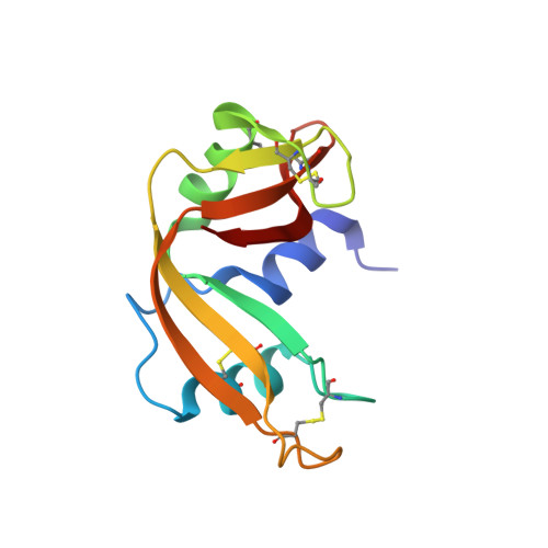
    5NA9

  • PubMed Abstract: 

    Cisplatin (CDDP) can form interprotein cross-links, leading to the formation of platinated oligomers. A dimer, a trimer and higher oligomers of bovine pancreatic ribonuclease (RNase A) obtained upon reaction with CDDP in 1:10 protein to metal ratio at 37°C have been previously characterized. Here, we verify the ability of carboplatin and oxaliplatin to induce RNase A oligomerization under the same experimental conditions. The amount of formed RNase A oligomers was compared with that obtained in the reaction of the protein with CDDP. Among the three anticancer agents, CDDP is the most reactive and the most effective in inhibiting the ribonucleolytic activity of the protein. Oxaliplatin is the least potent oligomerization agent. Biophysical characterizations of structure and stability of platinated dimers formed in the presence of carboplatin and oxaliplatin suggest that they have a similar thermal stability and are more prone to dissociation than the corresponding dimer obtained with CDDP. Oligomers obtained in the presence of carboplatin are the most active. X-ray structures of the monomeric adducts that RNase A forms with the three drugs provide a rational basis to explain the different effects of the three anticancer agents on enzymatic activity and protein aggregation. Although platinated oligomers of RNase A formed upon reaction with CDDP, carboplatin and oxaliplatin retain a residual ribonuclease activity, they do not show cytotoxic action, suggesting that protein aggregation processes induced by Pt-based drugs can represent a collateral drawback, which affects the functional state of protein targets and reduces the efficacy of Pt-based drug treatment.


  • Organizational Affiliation
    • Department of Chemical Sciences, University of Naples Federico II, via Cintia, I-80126 Naples, Italy. Electronic address: delia.picone@unina.it.

Macromolecule Content 

  • Total Structure Weight: 14.93 kDa 
  • Atom Count: 1,193 
  • Modeled Residue Count: 124 
  • Deposited Residue Count: 124 
  • Unique protein chains: 1

Macromolecules

Find similar proteins by:|  3D Structure
Entity ID: 1
MoleculeChains  Sequence LengthOrganismDetailsImage
Ribonuclease pancreatic124Bos taurusMutation(s): 0 
EC: 3.1.27.5 (PDB Primary Data), 4.6.1.18 (UniProt)
UniProt
Find proteins for P61823 (Bos taurus)
Explore P61823 
Go to UniProtKB:  P61823
Entity Groups
Sequence Clusters30% Identity50% Identity70% Identity90% Identity95% Identity100% Identity
UniProt GroupP61823
Sequence Annotations
Expand
Reference Sequence

Small Molecules

Ligands 3 Unique
IDChains Name / Formula / InChI Key2D Diagram3D Interactions
QPT

Query on QPT


Download:Ideal Coordinates CCD File
B [auth A]carboplatin
C6 H12 N2 O4 Pt
BHKICZDKIIDMNR-UHFFFAOYSA-L
CS

Query on CS


Download:Ideal Coordinates CCD File
C [auth A],
D [auth A],
E [auth A],
F [auth A]
CESIUM ION
Cs
NCMHKCKGHRPLCM-UHFFFAOYSA-N
CL

Query on CL


Download:Ideal Coordinates CCD File
G [auth A]
H [auth A]
I [auth A]
J [auth A]
K [auth A]
G [auth A],
H [auth A],
I [auth A],
J [auth A],
K [auth A],
L [auth A],
M [auth A],
N [auth A],
O [auth A]
CHLORIDE ION
Cl
VEXZGXHMUGYJMC-UHFFFAOYSA-M

Experimental Data & Validation

Experimental Data

  • Method: X-RAY DIFFRACTION
  • Resolution: 2.07 Å
  • R-Value Free:  0.244 (Depositor), 0.249 (DCC) 
  • R-Value Work:  0.178 (Depositor), 0.186 (DCC) 
  • R-Value Observed: 0.182 (Depositor) 
Space Group: P 32 2 1
Unit Cell:
Length ( Å )Angle ( ˚ )
a = 64.168α = 90
b = 64.168β = 90
c = 64.271γ = 120
Software Package:
Software NamePurpose
REFMACrefinement
HKL-2000data reduction
HKL-2000data scaling
PHASERphasing

Structure Validation

View Full Validation Report


Entry History 

Deposition Data

Revision History  (Full details and data files)

  • Version 1.0: 2017-08-16
    Type: Initial release
  • Version 1.1: 2024-01-17
    Changes: Advisory, Data collection, Database references, Derived calculations, Refinement description
  • Version 1.2: 2024-10-16
    Changes: Structure summary