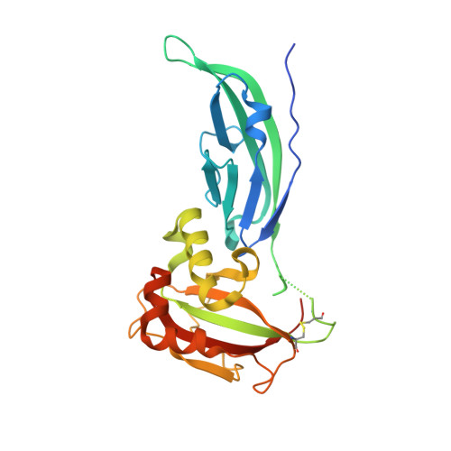
The effect of the pathological V72I, D109N and T190M missense mutations on the molecular structure of alpha-dystroglycan.
Covaceuszach, S., Bozzi, M., Bigotti, M.G., Sciandra, F., Konarev, P.V., Brancaccio, A., Cassetta, A.(2017) PLoS One 12: e0186110-e0186110
- PubMed: 29036200
- DOI: https://doi.org/10.1371/journal.pone.0186110
- Primary Citation of Related Structures:
5N30, 5N4H - PubMed Abstract:
Dystroglycan (DG) is a highly glycosylated protein complex that links the cytoskeleton with the extracellular matrix, mediating fundamental physiological functions such as mechanical stability of tissues, matrix organization and cell polarity. A crucial role in the glycosylation of the DG α subunit is played by its own N-terminal region that is required by the glycosyltransferase LARGE. Alteration in this O-glycosylation deeply impairs the high affinity binding to other extracellular matrix proteins such as laminins. Recently, three missense mutations in the gene encoding DG, mapped in the α-DG N-terminal region, were found to be responsible for hypoglycosylated states, causing congenital diseases of different severity referred as primary dystroglycanopaties.To gain insight on the molecular basis of these disorders, we investigated the crystallographic and solution structures of these pathological point mutants, namely V72I, D109N and T190M. Small Angle X-ray Scattering analysis reveals that these mutations affect the structures in solution, altering the distribution between compact and more elongated conformations. These results, supported by biochemical and biophysical assays, point to an altered structural flexibility of the mutant α-DG N-terminal region that may have repercussions on its interaction with LARGE and/or other DG-modifying enzymes, eventually reducing their catalytic efficiency.
- Istituto di Cristallografia-CNR, Trieste Outstation, Trieste, Italy.
Organizational Affiliation:


