5N1T | pdb_00005n1t

Crystal structure of complex between flavocytochrome c and copper chaperone CopC from T. paradoxus


Experimental Data Snapshot

  • Method: X-RAY DIFFRACTION
  • Resolution: 2.60 Å
  • R-Value Free: 
    0.257 (Depositor), 0.260 (DCC) 
  • R-Value Work: 
    0.186 (Depositor), 0.190 (DCC) 
  • R-Value Observed: 
    0.189 (Depositor) 

Starting Model: experimental
View more details

wwPDB Validation   3D Report Full Report


Ligand Structure Quality Assessment 

Created with Raphaël 2.3.0Worse 01 BetterLigand structure goodness of fit to experimental dataBest fitted FADClick on this verticalbar to view detailsBest fitted HECClick on this verticalbar to view details

This is version 1.3 of the entry. See complete history


Literature

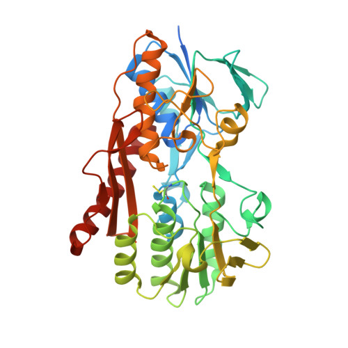
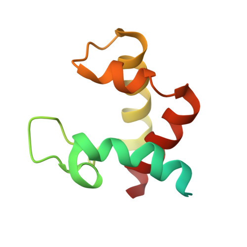
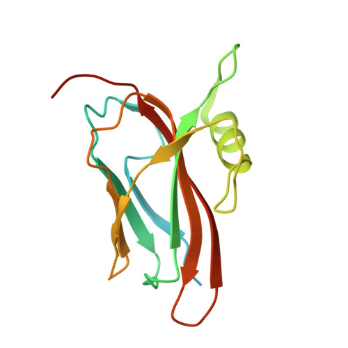
Structure of the flavocytochrome c sulfide dehydrogenase associated with the copper-binding protein CopC from the haloalkaliphilic sulfur-oxidizing bacterium Thioalkalivibrio paradoxusARh 1.

Osipov, E.M.Lilina, A.V.Tsallagov, S.I.Safonova, T.N.Sorokin, D.Y.Tikhonova, T.V.Popov, V.O.

(2018) Acta Crystallogr D Struct Biol 74: 632-642

  • DOI: https://doi.org/10.1107/S2059798318005648
  • Primary Citation of Related Structures:  
    5N1T

  • PubMed Abstract: 

    Flavocytochrome c sulfide dehydrogenase from Thioalkalivibrio paradoxus (TpFCC) is a heterodimeric protein consisting of flavin- and monohaem c-binding subunits. TpFCC was co-purified and co-crystallized with the dimeric copper-binding protein TpCopC. The structure of the TpFCC-(TpCopC) 2 complex was determined by X-ray diffraction at 2.6 Å resolution. The flavin-binding subunit of TpFCC is structurally similar to those determined previously, and the structure of the haem-binding subunit is similar to that of the N-terminal domain of dihaem FCCs. According to classification based on amino-acid sequence, TpCopC belongs to a high-affinity CopC subfamily characterized by the presence of a conserved His1-Xxx-His3 motif at the N-terminus. Apparently, a unique α-helix which is present in each monomer of TpCopC at the interface with TpFCC plays a key role in complex formation. The structure of the copper-binding site in TpCopC is similar to those in other known CopC structures. His3 is not involved in binding to the copper ion and is 6-7 Å away from this ion. Therefore, the His1-Xxx-His3 motif cannot be considered to be a key factor in the high affinity of CopC for copper(II) ions. It is suggested that the TpFCC-(TpCopC) 2 heterotetramer may be a component of a large periplasmic complex that is responsible for thiocyanate metabolism.


  • Organizational Affiliation

    Research Center of Biotechnology of the Russian Academy of Sciences, 33 Leninsky Avenue, Building 2, Moscow 119071, Russian Federation.


Macromolecules
Find similar proteins by:  (by identity cutoff)  |  3D Structure
Entity ID: 1
MoleculeChains Sequence LengthOrganismDetailsImage
flavin-binding subunit of sulfide dehydrogenase429Thioalkalivibrio paradoxus ARh 1Mutation(s): 0 
UniProt
Find proteins for W0DP88 (Thioalkalivibrio paradoxus ARh 1)
Explore W0DP88 
Go to UniProtKB:  W0DP88
Entity Groups  
Sequence Clusters30% Identity50% Identity70% Identity90% Identity95% Identity100% Identity
UniProt GroupW0DP88
Sequence Annotations
Expand
  • Reference Sequence
Find similar proteins by:  (by identity cutoff)  |  3D Structure
Entity ID: 2
MoleculeChains Sequence LengthOrganismDetailsImage
Cytochrome C102Thioalkalivibrio paradoxus ARh 1Mutation(s): 0 
UniProt
Find proteins for W0DKT4 (Thioalkalivibrio paradoxus ARh 1)
Explore W0DKT4 
Go to UniProtKB:  W0DKT4
Entity Groups  
Sequence Clusters30% Identity50% Identity70% Identity90% Identity95% Identity100% Identity
UniProt GroupW0DKT4
Sequence Annotations
Expand
  • Reference Sequence
Find similar proteins by:  (by identity cutoff)  |  3D Structure
Entity ID: 3
MoleculeChains Sequence LengthOrganismDetailsImage
CopCC [auth M],
D [auth W]
160Thioalkalivibrio paradoxus ARh 1Mutation(s): 0 
UniProt
Find proteins for W0DSL1 (Thioalkalivibrio paradoxus ARh 1)
Explore W0DSL1 
Go to UniProtKB:  W0DSL1
Entity Groups  
Sequence Clusters30% Identity50% Identity70% Identity90% Identity95% Identity100% Identity
UniProt GroupW0DSL1
Sequence Annotations
Expand
  • Reference Sequence
Experimental Data & Validation

Experimental Data

  • Method: X-RAY DIFFRACTION
  • Resolution: 2.60 Å
  • R-Value Free:  0.257 (Depositor), 0.260 (DCC) 
  • R-Value Work:  0.186 (Depositor), 0.190 (DCC) 
  • R-Value Observed: 0.189 (Depositor) 
Space Group: P 21 21 21
Unit Cell:
Length ( Å )Angle ( ˚ )
a = 39.53α = 90
b = 138.4β = 90
c = 155.81γ = 90
Software Package:
Software NamePurpose
REFMACrefinement
XDSdata reduction
XSCALEdata scaling
MOLREPphasing

Structure Validation

View Full Validation Report


Ligand Structure Quality Assessment 

Created with Raphaël 2.3.0Worse 01 BetterLigand structure goodness of fit to experimental dataBest fitted FADClick on this verticalbar to view detailsBest fitted HECClick on this verticalbar to view details

Entry History & Funding Information

Deposition Data


Funding OrganizationLocationGrant Number
RNFRussian Federation14-24-00172

Revision History  (Full details and data files)

  • Version 1.0: 2018-02-28
    Type: Initial release
  • Version 1.1: 2018-07-11
    Changes: Data collection, Database references
  • Version 1.2: 2019-08-14
    Changes: Data collection
  • Version 1.3: 2024-01-17
    Changes: Data collection, Database references, Derived calculations, Refinement description