5N1A | pdb_00005n1a

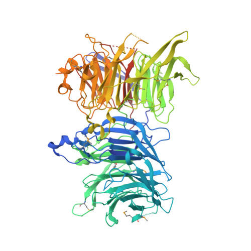
Crystal structure of Utp4 from Chaetomium thermophilum


Experimental Data Snapshot

  • Method: X-RAY DIFFRACTION
  • Resolution: 2.15 Å
  • R-Value Free: 
    0.220 (Depositor), 0.219 (DCC) 
  • R-Value Work: 
    0.183 (Depositor), 0.182 (DCC) 
  • R-Value Observed: 
    0.185 (Depositor) 

wwPDB Validation 3D Report Full Report

Validation slider image for 5N1A

This is version 1.2 of the entry. See complete history. 

Literature

Structural basis for 5'-ETS recognition by Utp4 at the early stages of ribosome biogenesis.

Calvino, F.R., Kornprobst, M., Schermann, G., Birkle, F., Wild, K., Fischer, T., Hurt, E., Ahmed, Y.L., Sinning, I.

(2017) PLoS One 12: e0178752-e0178752

  • DOI: https://doi.org/10.1371/journal.pone.0178752
  • Primary Citation Related Structures: 
    5N1A

  • PubMed Abstract: 

    Eukaryotic ribosome biogenesis begins with the co-transcriptional assembly of the 90S pre-ribosome. The 'U three protein' (UTP) complexes and snoRNP particles arrange around the nascent pre-ribosomal RNA chaperoning its folding and further maturation. The earliest event in this hierarchical process is the binding of the UTP-A complex to the 5'-end of the pre-ribosomal RNA (5'-ETS). This oligomeric complex predominantly consists of β-propeller and α-solenoidal proteins. Here we present the structure of the Utp4 subunit from the thermophilic fungus Chaetomium thermophilum at 2.15 Å resolution and analyze its function by UV RNA-crosslinking (CRAC) and in context of a recent cryo-EM structure of the 90S pre-ribosome. Utp4 consists of two orthogonal and highly basic β-propellers that perfectly fit the EM-data. The Utp4 structure highlights an unusual Velcro-closure of its C-terminal β-propeller as relevant for protein integrity and potentially Utp8 recognition in the context of the pre-ribosome. We provide a first model of the 5'-ETS RNA from the internally hidden 5'-end up to the region that hybridizes to the 3'-hinge sequence of U3 snoRNA and validate a specific Utp4/5'-ETS interaction by CRAC analysis.


  • Organizational Affiliation: 
    • Heidelberg University Biochemistry Center (BZH), Im Neuenheimer Feld 328, Heidelberg, Germany.

Macromolecule Content 

  • Total Structure Weight: 202.94 kDa 
  • Atom Count: 11,717 
  • Modeled Residue Count: 1,409 
  • Deposited Residue Count: 1,816 
  • Unique protein chains: 1

Macromolecules

Find similar proteins by:|  3D Structure
Entity ID: 1
MoleculeChains  Sequence LengthOrganismDetailsImage
utp4
A, B
908Thermochaetoides thermophila DSM 1495Mutation(s): 0 
Gene Names: CTHT_0058380
UniProt
Find proteins for G0SCT7 (Thermochaetoides thermophila (strain DSM 1495 / CBS 144.50 / DAOM 24625 / IMI 039719))
Explore G0SCT7 
Go to UniProtKB:  G0SCT7
Entity Groups
Sequence Clusters30% Identity50% Identity70% Identity90% Identity95% Identity100% Identity
UniProt GroupG0SCT7
Sequence Annotations
Expand
Reference Sequence

Small Molecules

Modified Residues  1 Unique
IDChains TypeFormula2D DiagramParent
MSE
Query on MSE
A, B
L-PEPTIDE LINKINGC5 H11 N O2 SeMET

Experimental Data & Validation

Experimental Data

  • Method: X-RAY DIFFRACTION
  • Resolution: 2.15 Å
  • R-Value Free:  0.220 (Depositor), 0.219 (DCC) 
  • R-Value Work:  0.183 (Depositor), 0.182 (DCC) 
  • R-Value Observed: 0.185 (Depositor) 
Space Group: C 1 2 1
Unit Cell:
Length ( Å )Angle ( ˚ )
a = 203.027α = 90
b = 81.593β = 110.55
c = 112.3γ = 90
Software Package:
Software NamePurpose
PHENIXrefinement
Aimlessdata scaling
PDB_EXTRACTdata extraction
XDSdata reduction
PHENIXphasing

Structure Validation

View Full Validation Report


Entry History 

& Funding Information

Deposition Data


Funding OrganizationLocationGrant Number
German Research FoundationGermany--

Revision History  (Full details and data files)

  • Version 1.0: 2017-06-14
    Type: Initial release
  • Version 1.1: 2019-10-16
    Changes: Author supporting evidence, Data collection
  • Version 1.2: 2024-11-06
    Changes: Data collection, Database references, Refinement description, Structure summary