5MRV

Crystal structure of human carboxypeptidase O in complex with NvCI


Experimental Data Snapshot

  • Method: X-RAY DIFFRACTION
  • Resolution: 1.85 Å
  • R-Value Free: 0.208 
  • R-Value Work: 0.185 
  • R-Value Observed: 0.186 

Starting Model: experimental
View more details

wwPDB Validation   3D Report Full Report


Ligand Structure Quality Assessment 


This is version 1.3 of the entry. See complete history


Literature

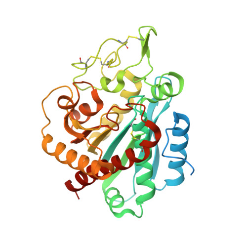
Crystal structure and mechanism of human carboxypeptidase O: Insights into its specific activity for acidic residues.

Garcia-Guerrero, M.C.Garcia-Pardo, J.Berenguer, E.Fernandez-Alvarez, R.Barfi, G.B.Lyons, P.J.Aviles, F.X.Huber, R.Lorenzo, J.Reverter, D.

(2018) Proc Natl Acad Sci U S A 115: E3932-E3939

  • DOI: https://doi.org/10.1073/pnas.1803685115
  • Primary Citation of Related Structures:  
    5MRV

  • PubMed Abstract: 

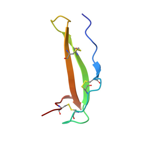
    Human metallocarboxypeptidase O (hCPO) is a recently discovered digestive enzyme localized to the apical membrane of intestinal epithelial cells. Unlike pancreatic metallocarboxypeptidases, hCPO is glycosylated and produced as an active enzyme with distinctive substrate specificity toward C-terminal (C-t) acidic residues. Here we present the crystal structure of hCPO at 1.85-Å resolution, both alone and in complex with a carboxypeptidase inhibitor (NvCI) from the marine snail Nerita versicolor The structure provides detailed information regarding determinants of enzyme specificity, in particular Arg275, placed at the bottom of the substrate-binding pocket. This residue, located at "canonical" position 255, where it is Ile in human pancreatic carboxypeptidases A1 (hCPA1) and A2 (hCPA2) and Asp in B (hCPB), plays a dominant role in determining the preference of hCPO for acidic C-t residues. Site-directed mutagenesis to Asp and Ala changes the specificity to C-t basic and hydrophobic residues, respectively. The single-site mutants thus faithfully mimic the enzymatic properties of CPB and CPA, respectively. hCPO also shows a preference for Glu over Asp, probably as a consequence of a tighter fitting of the Glu side chain in its S1' substrate-binding pocket. This unique preference of hCPO, together with hCPA1, hCPA2, and hCPB, completes the array of C-t cleavages enabling the digestion of the dietary proteins within the intestine. Finally, in addition to activity toward small synthetic substrates and peptides, hCPO can also trim C-t extensions of proteins, such as epidermal growth factor, suggesting a role in the maturation and degradation of growth factors and bioactive peptides.


  • Organizational Affiliation

    Institute for Biotechnology and Biomedicine, Universitat Autonoma de Barcelona, 08193 Bellaterra, Barcelona, Spain.


Macromolecules
Find similar proteins by:  (by identity cutoff)  |  3D Structure
Entity ID: 1
MoleculeChains Sequence LengthOrganismDetailsImage
Carboxypeptidase O
A, B
355Homo sapiensMutation(s): 2 
Gene Names: CPO
EC: 3.4.17
UniProt & NIH Common Fund Data Resources
Find proteins for Q8IVL8 (Homo sapiens)
Explore Q8IVL8 
Go to UniProtKB:  Q8IVL8
PHAROS:  Q8IVL8
GTEx:  ENSG00000144410 
Entity Groups  
Sequence Clusters30% Identity50% Identity70% Identity90% Identity95% Identity100% Identity
UniProt GroupQ8IVL8
Glycosylation
Glycosylation Sites: 2Go to GlyGen: Q8IVL8-1
Sequence Annotations
Expand
  • Reference Sequence
Find similar proteins by:  (by identity cutoff)  |  3D Structure
Entity ID: 2
MoleculeChains Sequence LengthOrganismDetailsImage
Metallocarboxypeptidase inhibitor53Nerita versicolorMutation(s): 0 
UniProt
Find proteins for P86912 (Nerita versicolor)
Explore P86912 
Go to UniProtKB:  P86912
Entity Groups  
Sequence Clusters30% Identity50% Identity70% Identity90% Identity95% Identity100% Identity
UniProt GroupP86912
Sequence Annotations
Expand
  • Reference Sequence
Experimental Data & Validation

Experimental Data

  • Method: X-RAY DIFFRACTION
  • Resolution: 1.85 Å
  • R-Value Free: 0.208 
  • R-Value Work: 0.185 
  • R-Value Observed: 0.186 
  • Space Group: C 1 2 1
Unit Cell:
Length ( Å )Angle ( ˚ )
a = 150.114α = 90
b = 72.144β = 94.66
c = 90.187γ = 90
Software Package:
Software NamePurpose
PHENIXrefinement
XDSdata reduction
Aimlessdata scaling
PHASERphasing

Structure Validation

View Full Validation Report


Ligand Structure Quality Assessment 


Entry History 

Deposition Data

Revision History  (Full details and data files)

  • Version 1.0: 2018-01-17
    Type: Initial release
  • Version 1.1: 2018-05-16
    Changes: Data collection, Database references
  • Version 1.2: 2020-07-29
    Type: Remediation
    Reason: Carbohydrate remediation
    Changes: Data collection, Derived calculations, Structure summary
  • Version 1.3: 2024-01-17
    Changes: Data collection, Database references, Refinement description, Structure summary