Phosphorylation decelerates conformational dynamics in bacterial translation elongation factors.
Talavera, A., Hendrix, J., Versees, W., Jurenas, D., Van Nerom, K., Vandenberk, N., Singh, R.K., Konijnenberg, A., De Gieter, S., Castro-Roa, D., Barth, A., De Greve, H., Sobott, F., Hofkens, J., Zenkin, N., Loris, R., Garcia-Pino, A.(2018) Sci Adv 4: eaap9714-eaap9714
- PubMed: 29546243 Search on PubMedSearch on PubMed Central
- DOI: https://doi.org/10.1126/sciadv.aap9714
- Primary Citation Related Structures:
5MI3, 5MI8, 5MI9 - PubMed Abstract:
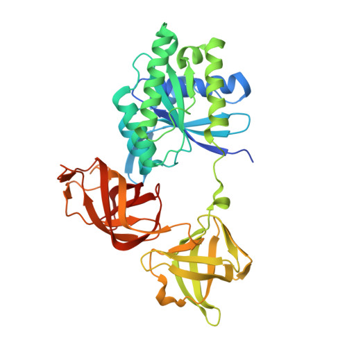
Bacterial protein synthesis is intricately connected to metabolic rate. One of the ways in which bacteria respond to environmental stress is through posttranslational modifications of translation factors. Translation elongation factor Tu (EF-Tu) is methylated and phosphorylated in response to nutrient starvation upon entering stationary phase, and its phosphorylation is a crucial step in the pathway toward sporulation. We analyze how phosphorylation leads to inactivation of Escherichia coli EF-Tu. We provide structural and biophysical evidence that phosphorylation of EF-Tu at T382 acts as an efficient switch that turns off protein synthesis by decoupling nucleotide binding from the EF-Tu conformational cycle. Direct modifications of the EF-Tu switch I region or modifications in other regions stabilizing the β-hairpin state of switch I result in an effective allosteric trap that restricts the normal dynamics of EF-Tu and enables the evasion of the control exerted by nucleotides on G proteins. These results highlight stabilization of a phosphorylation-induced conformational trap as an essential mechanism for phosphoregulation of bacterial translation and metabolism. We propose that this mechanism may lead to the multisite phosphorylation state observed during dormancy and stationary phase.
- Structural Biology Brussels, Department of Bio-engineering Sciences, Vrije Universiteit Brussel, Brussels, Belgium.
Organizational Affiliation: