5MGB

Crystal Structure of Rat Peroxisomal Multifunctional enzyme Type-1 (RPMFE1) Complexed with Acetoacetyl-CoA and NAD


Experimental Data Snapshot

  • Method: X-RAY DIFFRACTION
  • Resolution: 2.80 Å
  • R-Value Free: 0.261 
  • R-Value Work: 0.209 
  • R-Value Observed: 0.212 

wwPDB Validation   3D Report Full Report


Ligand Structure Quality Assessment 


This is version 1.5 of the entry. See complete history


Literature

Structural enzymology comparisons of multifunctional enzyme, type-1 (MFE1): the flexibility of its dehydrogenase part.

Kasaragod, P.Midekessa, G.B.Sridhar, S.Schmitz, W.Kiema, T.R.Hiltunen, J.K.Wierenga, R.K.

(2017) FEBS Open Bio 7: 1830-1842

  • DOI: https://doi.org/10.1002/2211-5463.12337
  • Primary Citation of Related Structures:  
    5MGB

  • PubMed Abstract: 

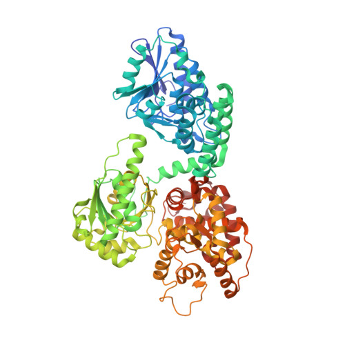
    Multifunctional enzyme, type-1 (MFE1) is a monomeric enzyme with a 2E-enoyl-CoA hydratase and a 3S-hydroxyacyl-CoA dehydrogenase (HAD) active site. Enzyme kinetic data of rat peroxisomal MFE1 show that the catalytic efficiencies for converting the short-chain substrate 2E-butenoyl-CoA into acetoacetyl-CoA are much lower when compared with those of the homologous monofunctional enzymes. The mode of binding of acetoacetyl-CoA (to the hydratase active site) and the very similar mode of binding of NAD + and NADH (to the HAD part) are described and compared with those of their monofunctional counterparts. Structural comparisons suggest that the conformational flexibility of the HAD and hydratase parts of MFE1 are correlated. The possible importance of the conformational flexibility of MFE1 for its biocatalytic properties is discussed.


  • Organizational Affiliation

    Biocenter Oulu and Faculty of Biochemistry and Molecular Medicine University of Oulu Finland.


Macromolecules
Find similar proteins by:  (by identity cutoff)  |  3D Structure
Entity ID: 1
MoleculeChains Sequence LengthOrganismDetailsImage
Peroxisomal bifunctional enzyme
A, B
742Rattus norvegicusMutation(s): 0 
Gene Names: Ehhadh
EC: 4.2.1.17 (PDB Primary Data), 5.3.3.8 (PDB Primary Data), 1.1.1.35 (PDB Primary Data)
UniProt
Find proteins for P07896 (Rattus norvegicus)
Explore P07896 
Go to UniProtKB:  P07896
Entity Groups  
Sequence Clusters30% Identity50% Identity70% Identity90% Identity95% Identity100% Identity
UniProt GroupP07896
Sequence Annotations
Expand
  • Reference Sequence
Small Molecules
Ligands 4 Unique
IDChains Name / Formula / InChI Key2D Diagram3D Interactions
CAA
Query on CAA

Download Ideal Coordinates CCD File 
F [auth A],
K [auth B]
ACETOACETYL-COENZYME A
C25 H40 N7 O18 P3 S
OJFDKHTZOUZBOS-CITAKDKDSA-N
NAD
Query on NAD

Download Ideal Coordinates CCD File 
G [auth A],
L [auth B]
NICOTINAMIDE-ADENINE-DINUCLEOTIDE
C21 H27 N7 O14 P2
BAWFJGJZGIEFAR-NNYOXOHSSA-N
SO4
Query on SO4

Download Ideal Coordinates CCD File 
C [auth A],
D [auth A],
E [auth A],
J [auth B]
SULFATE ION
O4 S
QAOWNCQODCNURD-UHFFFAOYSA-L
GOL
Query on GOL

Download Ideal Coordinates CCD File 
H [auth A],
I [auth A]
GLYCEROL
C3 H8 O3
PEDCQBHIVMGVHV-UHFFFAOYSA-N
Experimental Data & Validation

Experimental Data

  • Method: X-RAY DIFFRACTION
  • Resolution: 2.80 Å
  • R-Value Free: 0.261 
  • R-Value Work: 0.209 
  • R-Value Observed: 0.212 
  • Space Group: P 21 21 21
Unit Cell:
Length ( Å )Angle ( ˚ )
a = 65.23α = 90
b = 125.82β = 90
c = 223.9γ = 90
Software Package:
Software NamePurpose
REFMACrefinement
MOSFLMdata reduction
SCALAdata scaling
REFMACphasing

Structure Validation

View Full Validation Report


Ligand Structure Quality Assessment 


Entry History 

Deposition Data

Revision History  (Full details and data files)

  • Version 1.0: 2016-12-21
    Type: Initial release
  • Version 1.1: 2017-03-08
    Changes: Other
  • Version 1.2: 2017-08-09
    Changes: Structure summary
  • Version 1.3: 2017-11-15
    Changes: Database references
  • Version 1.4: 2017-12-20
    Changes: Database references
  • Version 1.5: 2024-01-17
    Changes: Data collection, Database references, Refinement description