5MEW | pdb_00005mew

The study of the X-ray induced enzymatic reduction of molecular oxygen to water for laccase from Steccherinum murashkinskyi. Second structure of the series with total exposition time 33 min.


Experimental Data Snapshot

  • Method: X-RAY DIFFRACTION
  • Resolution: 1.35 Å
  • R-Value Free: 
    0.152 (Depositor), 0.154 (DCC) 
  • R-Value Work: 
    0.132 (Depositor), 0.133 (DCC) 
  • R-Value Observed: 
    0.133 (Depositor) 

Starting Model: experimental
View more details

wwPDB Validation 3D Report Full Report

Validation slider image for 5MEW

This is version 2.2 of the entry. See complete history

Literature

Structural study of the X-ray-induced enzymatic reduction of molecular oxygen to water by Steccherinum murashkinskyi laccase: insights into the reaction mechanism.

Polyakov, K.M.Gavryushov, S.Ivanova, S.Fedorova, T.V.Glazunova, O.A.Popov, A.N.Koroleva, O.V.

(2017) Acta Crystallogr D Struct Biol 73: 388-401

  • DOI: https://doi.org/10.1107/S2059798317003667
  • Primary Citation Related Structures: 
    5MEJ, 5MEW, 5MHU, 5MHV, 5MHW, 5MHX, 5MHY, 5MHZ, 5MI1, 5MI2, 5MIA, 5MIB, 5MIC, 5MID, 5MIE, 5MIG

  • PubMed Abstract: 

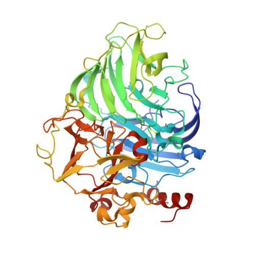
    The laccase from Steccherinum murashkinskyi is a member of the large family of multicopper oxidases that catalyze the oxidation of a wide range of organic and inorganic substrates, accompanied by the reduction of dioxygen to water. The reducing properties of X-ray radiation and the high quality of the laccase crystals allow the study of the catalytic reduction of dioxygen to water directly in a crystal. A series of diffraction data sets with increasing absorbed radiation dose were collected from a single crystal of Steccherinum murashkinskyi laccase at 1.35 Å resolution. Changes in the active-site structure associated with the reduction of molecular oxygen to water on increasing the absorbed dose of ionizing radiation were detected. The structures in the series are mixtures of different states of the enzyme-substrate complex. Nevertheless, it was possible to interpret these structures as complexes of various oxygen ligands with copper ions in different oxidation states. The results allowed the mechanism of oxygen reduction catalyzed by laccases to be refined.


  • Organizational Affiliation
    • Engelhardt Institute of Molecular Biology, Russian Academy of Sciences, Vavilova Str. 32, Moscow 119991, Russian Federation.

Macromolecule Content 

  • Total Structure Weight: 55.09 kDa 
  • Atom Count: 4,748 
  • Modeled Residue Count: 499 
  • Deposited Residue Count: 499 
  • Unique protein chains: 1

Macromolecules

Find similar proteins by:|  3D Structure
Entity ID: 1
MoleculeChains  Sequence LengthOrganismDetailsImage
Laccase 2499Metuloidea murashkinskyiMutation(s): 0 
EC: 1.10.3.2
UniProt
Find proteins for I1VE66 (Metuloidea murashkinskyi)
Explore I1VE66 
Go to UniProtKB:  I1VE66
Entity Groups
Sequence Clusters30% Identity50% Identity70% Identity90% Identity95% Identity100% Identity
UniProt GroupI1VE66
Glycosylation
Glycosylation Sites: 2
Sequence Annotations
Expand
Reference Sequence

Oligosaccharides

Help  
Entity ID: 2
MoleculeChains Length2D Diagram GlycosylationD Interactions
beta-D-mannopyranose-(1-4)-2-acetamido-2-deoxy-beta-D-glucopyranose-(1-4)-2-acetamido-2-deoxy-beta-D-glucopyranose
B, C
3N-Glycosylation
Glycosylation Resources
GlyTouCan: G15407YE
GlyCosmos: G15407YE
GlyGen: G15407YE

Small Molecules

Ligands 4 Unique
IDChains Name / Formula / InChI Key2D Diagram3D Interactions
CIT

Query on CIT


Download:Ideal Coordinates CCD File
J [auth A]CITRIC ACID
C6 H8 O7
KRKNYBCHXYNGOX-UHFFFAOYSA-N
CU

Query on CU


Download:Ideal Coordinates CCD File
D [auth A],
E [auth A],
F [auth A],
G [auth A]
COPPER (II) ION
Cu
JPVYNHNXODAKFH-UHFFFAOYSA-N
OXY

Query on OXY


Download:Ideal Coordinates CCD File
H [auth A],
I [auth A]
OXYGEN MOLECULE
O2
MYMOFIZGZYHOMD-UHFFFAOYSA-N
NA

Query on NA


Download:Ideal Coordinates CCD File
K [auth A]SODIUM ION
Na
FKNQFGJONOIPTF-UHFFFAOYSA-N

Experimental Data & Validation

Experimental Data

  • Method: X-RAY DIFFRACTION
  • Resolution: 1.35 Å
  • R-Value Free:  0.152 (Depositor), 0.154 (DCC) 
  • R-Value Work:  0.132 (Depositor), 0.133 (DCC) 
  • R-Value Observed: 0.133 (Depositor) 
Space Group: P 21 21 21
Unit Cell:
Length ( Å )Angle ( ˚ )
a = 56.38α = 90
b = 84.4β = 90
c = 112.49γ = 90
Software Package:
Software NamePurpose
REFMACrefinement
XSCALEdata scaling
XDSdata reduction

Structure Validation

View Full Validation Report


Entry History 

& Funding Information

Deposition Data


Funding OrganizationLocationGrant Number
RFBRRussian Federation16-04-00552

Revision History  (Full details and data files)

  • Version 1.0: 2017-05-10
    Type: Initial release
  • Version 1.1: 2017-05-24
    Changes: Database references
  • Version 1.2: 2018-01-31
    Changes: Database references
  • Version 1.3: 2019-07-17
    Changes: Data collection
  • Version 2.0: 2020-07-29
    Type: Remediation
    Reason: Carbohydrate remediation
    Changes: Advisory, Atomic model, Data collection, Derived calculations, Structure summary
  • Version 2.1: 2024-01-17
    Changes: Data collection, Database references, Refinement description, Structure summary
  • Version 2.2: 2024-10-16
    Changes: Structure summary