5ME5 | pdb_00005me5

Crystal Structure of eiF4E from C. melo bound to a eIF4G peptide


Experimental Data Snapshot

  • Method: X-RAY DIFFRACTION
  • Resolution: 1.90 Å
  • R-Value Free: 
    0.213 (Depositor), 0.217 (DCC) 
  • R-Value Work: 
    0.186 (Depositor), 0.187 (DCC) 
  • R-Value Observed: 
    0.188 (Depositor) 

wwPDB Validation 3D Report Full Report

Validation slider image for 5ME5

This is version 1.1 of the entry. See complete history

Literature

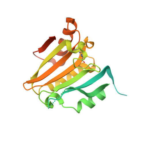
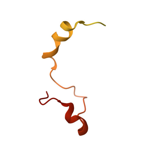
Structure of eIF4E in Complex with an eIF4G Peptide Supports a Universal Bipartite Binding Mode for Protein Translation.

Miras, M.Truniger, V.Silva, C.Verdaguer, N.Aranda, M.A.Querol-Audi, J.

(2017) Plant Physiol 174: 1476-1491

  • DOI: https://doi.org/10.1104/pp.17.00193
  • Primary Citation Related Structures: 
    5ME5, 5ME6, 5ME7

  • PubMed Abstract: 

    The association-dissociation of the cap-binding protein eukaryotic translation initiation factor 4E (eIF4E) with eIF4G is a key control step in eukaryotic translation. The paradigm on the eIF4E-eIF4G interaction states that eIF4G binds to the dorsal surface of eIF4E through a single canonical alpha-helical motif, while metazoan eIF4E-binding proteins (m4E-BPs) advantageously compete against eIF4G via bimodal interactions involving this canonical motif and a second noncanonical motif of the eIF4E surface. Metazoan eIF4Gs share this extended binding interface with m4E-BPs, with significant implications on the understanding of translation regulation and the design of therapeutic molecules. Here we show the high-resolution structure of melon ( Cucumis melo ) eIF4E in complex with a melon eIF4G peptide and propose the first eIF4E-eIF4G structural model for plants. Our structural data together with functional analyses demonstrate that plant eIF4G binds to eIF4E through both the canonical and noncanonical motifs, similarly to metazoan eIF4E-eIF4G complexes. As in the case of metazoan eIF4E-eIF4G, this may have very important practical implications, as plant eIF4E-eIF4G is also involved in a significant number of plant diseases. In light of our results, a universal eukaryotic bipartite mode of binding to eIF4E is proposed.


  • Organizational Affiliation
    • Centro de Edafología y Biología Aplicada del Segura (CEBAS), Consejo Superior de Investigaciones Científicas (CSIC), 30100 Espinardo, Murcia, Spain.

Macromolecule Content 

  • Total Structure Weight: 36.73 kDa 
  • Atom Count: 1,610 
  • Modeled Residue Count: 189 
  • Deposited Residue Count: 327 
  • Unique protein chains: 2

Macromolecules

Find similar proteins by:|  3D Structure
Entity ID: 1
MoleculeChains  Sequence LengthOrganismDetailsImage
Eukaryotic transcription initiation factor 4E237Cucumis meloMutation(s): 0 
Find similar proteins by:|  3D Structure
Entity ID: 2
MoleculeChains  Sequence LengthOrganismDetailsImage
eIF4G90Cucumis meloMutation(s): 0 
UniProt
Find proteins for A0A1S3C4H6 (Cucumis melo)
Explore A0A1S3C4H6 
Go to UniProtKB:  A0A1S3C4H6
Entity Groups
Sequence Clusters30% Identity50% Identity70% Identity90% Identity95% Identity100% Identity
UniProt GroupA0A1S3C4H6
Sequence Annotations
Expand
Reference Sequence

Experimental Data & Validation

Experimental Data

  • Method: X-RAY DIFFRACTION
  • Resolution: 1.90 Å
  • R-Value Free:  0.213 (Depositor), 0.217 (DCC) 
  • R-Value Work:  0.186 (Depositor), 0.187 (DCC) 
  • R-Value Observed: 0.188 (Depositor) 
Space Group: P 1 21 1
Unit Cell:
Length ( Å )Angle ( ˚ )
a = 38.094α = 90
b = 70.352β = 93.62
c = 42.798γ = 90
Software Package:
Software NamePurpose
PHENIXrefinement
SCALAdata scaling
XDSdata reduction
PDB_EXTRACTdata extraction
XSCALEdata scaling
PHASERphasing

Structure Validation

View Full Validation Report


Entry History 

Deposition Data

Revision History  (Full details and data files)

  • Version 1.0: 2017-08-23
    Type: Initial release
  • Version 1.1: 2024-11-13
    Changes: Data collection, Database references, Structure summary