5M4M | pdb_00005m4m

Application of Off-Rate Screening in the Identification of Novel Pan-Isoform Inhibitors of Pyruvate Dehydrogenase Kinase


Experimental Data Snapshot

  • Method: X-RAY DIFFRACTION
  • Resolution: 2.40 Å
  • R-Value Free: 
    0.262 (Depositor), 0.261 (DCC) 
  • R-Value Work: 
    0.217 (Depositor), 0.221 (DCC) 
  • R-Value Observed: 
    0.219 (Depositor) 

Starting Model: experimental
View more details

wwPDB Validation 3D Report Full Report

Validation slider image for 5M4M

Ligand Structure Quality Assessment 


This is version 1.2 of the entry. See complete history

Literature

Application of Off-Rate Screening in the Identification of Novel Pan-Isoform Inhibitors of Pyruvate Dehydrogenase Kinase.

Brough, P.A.Baker, L.Bedford, S.Brown, K.Chavda, S.Chell, V.D'Alessandro, J.Davies, N.G.Davis, B.Le Strat, L.Macias, A.T.Maddox, D.Mahon, P.C.Massey, A.J.Matassova, N.McKenna, S.Meissner, J.W.Moore, J.D.Murray, J.B.Northfield, C.J.Parry, C.Parsons, R.Roughley, S.D.Shaw, T.Simmonite, H.Stokes, S.Surgenor, A.Stefaniak, E.Robertson, A.Wang, Y.Webb, P.Whitehead, N.Wood, M.

(2017) J Med Chem 60: 2271-2286

  • DOI: https://doi.org/10.1021/acs.jmedchem.6b01478
  • Primary Citation Related Structures: 
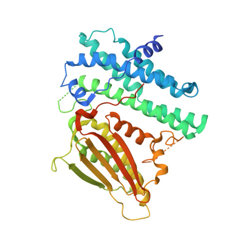
    5M4E, 5M4H, 5M4K, 5M4M, 5M4N, 5M4P

  • PubMed Abstract: 

    Libraries of nonpurified resorcinol amide derivatives were screened by surface plasmon resonance (SPR) to determine the binding dissociation constant (off-rate, k d ) for compounds binding to the pyruvate dehydrogenase kinase (PDHK) enzyme. Parallel off-rate measurements against HSP90 and application of structure-based drug design enabled rapid hit to lead progression in a program to identify pan-isoform ATP-competitive inhibitors of PDHK. Lead optimization identified selective sub-100-nM inhibitors of the enzyme which significantly reduced phosphorylation of the E1α subunit in the PC3 cancer cell line in vitro.


  • Organizational Affiliation
    • Vernalis (R&D) Ltd. , Granta Park, Great Abington, Cambridge CB21 6GB, U.K.

Macromolecule Content 

  • Total Structure Weight: 47.28 kDa 
  • Atom Count: 2,926 
  • Modeled Residue Count: 344 
  • Deposited Residue Count: 408 
  • Unique protein chains: 1

Macromolecules

Find similar proteins by:|  3D Structure
Entity ID: 1
MoleculeChains  Sequence LengthOrganismDetailsImage
[Pyruvate dehydrogenase (acetyl-transferring)] kinase isozyme 2, mitochondrial408Homo sapiensMutation(s): 0 
Gene Names: PDK2PDHK2
EC: 2.7.11.2
UniProt & NIH Common Fund Data Resources
Find proteins for Q15119 (Homo sapiens)
Explore Q15119 
Go to UniProtKB:  Q15119
PHAROS:  Q15119
GTEx:  ENSG00000005882 
Entity Groups
Sequence Clusters30% Identity50% Identity70% Identity90% Identity95% Identity100% Identity
UniProt GroupQ15119
Sequence Annotations
Expand
Reference Sequence

Small Molecules

Binding Affinity Annotations 
IDSourceBinding Affinity
TF3 BindingDB:  5M4M IC50: 2000 (nM) from 1 assay(s)

Experimental Data & Validation

Experimental Data

  • Method: X-RAY DIFFRACTION
  • Resolution: 2.40 Å
  • R-Value Free:  0.262 (Depositor), 0.261 (DCC) 
  • R-Value Work:  0.217 (Depositor), 0.221 (DCC) 
  • R-Value Observed: 0.219 (Depositor) 
Space Group: P 64
Unit Cell:
Length ( Å )Angle ( ˚ )
a = 108.651α = 90
b = 108.651β = 90
c = 83.657γ = 120
Software Package:
Software NamePurpose
REFMACrefinement
d*TREKdata reduction
d*TREKdata scaling
AMoREphasing

Structure Validation

View Full Validation Report


Ligand Structure Quality Assessment 


Entry History 

Deposition Data

Revision History  (Full details and data files)

  • Version 1.0: 2017-02-22
    Type: Initial release
  • Version 1.1: 2017-04-05
    Changes: Database references
  • Version 1.2: 2024-01-17
    Changes: Data collection, Database references, Refinement description