5M2H | pdb_00005m2h

Crystal structure of vancomycin-Zn(II)-citrate complex


Experimental Data Snapshot

  • Method: X-RAY DIFFRACTION
  • Resolution: 0.95 Å
  • R-Value Free: 
    0.118 (Depositor), 0.126 (DCC) 
  • R-Value Work: 
    0.105 (Depositor), 0.112 (DCC) 
  • R-Value Observed: 
    0.106 (Depositor) 

wwPDB Validation 3D Report Full Report

Validation slider image for 5M2H

This is version 3.1 of the entry. See complete history

Literature

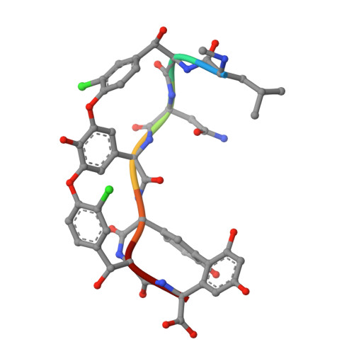
Zn(II) mediates vancomycin polymerization and potentiates its antibiotic activity against resistant bacteria.

Zarkan, A.Macklyne, H.R.Chirgadze, D.Y.Bond, A.D.Hesketh, A.R.Hong, H.J.

(2017) Sci Rep 7: 4893-4893

  • DOI: https://doi.org/10.1038/s41598-017-04868-2
  • Primary Citation Related Structures: 
    5M2H, 5M2K

  • PubMed Abstract: 

    Vancomycin is known to bind to Zn(II) and can induce a zinc starvation response in bacteria. Here we identify a novel polymerization of vancomycin dimers by structural analysis of vancomycin-Zn(II) crystals and fibre X-ray diffraction. Bioassays indicate that this structure is associated with an increased antibiotic activity against bacterial strains possessing high level vancomycin resistance mediated by the reprogramming of peptidoglycan biosynthesis to use precursors terminating in D-Ala-D-Lac in place of D-Ala-D-Ala. Polymerization occurs via interaction of Zn(II) with the N-terminal methylleucine group of vancomycin, and we show that the activity of other glycopeptide antibiotics with this feature can also be similarly augmented by Zn(II). Construction and analysis of a model strain predominantly using D-Ala-D-Lac precursors for peptidoglycan biosynthesis during normal growth supports the hypothesis that Zn(II) mediated vancomycin polymerization enhances the binding affinity towards these precursors.


  • Organizational Affiliation
    • Department of Biochemistry, University of Cambridge, Cambridge, CB2 1QW, UK.

Macromolecule Content 

  • Total Structure Weight: 3.68 kDa 
  • Atom Count: 320 
  • Modeled Residue Count: 14 
  • Deposited Residue Count: 14 
  • Unique protein chains: 1

Macromolecules

Find similar proteins by:  Sequence   |   3D Structure  
Entity ID: 1
MoleculeChains  Sequence LengthOrganismDetailsImage
Vancomycin
A, B
7Amycolatopsis orientalisMutation(s): 0 

Oligosaccharides

Help  
Entity ID: 2
MoleculeChains Length2D Diagram GlycosylationD Interactions
vancosamine-(1-2)-beta-D-glucopyranose
C, D
2N/A
Glycosylation Resources
GlyTouCan: G14263HU
GlyCosmos: G14263HU

Small Molecules

Modified Residues  1 Unique
IDChains TypeFormula2D DiagramParent
3FG
Query on 3FG
A, B
L-PEPTIDE LINKINGC8 H9 N O4

--

GHP
Query on GHP
A, B
D-PEPTIDE LINKINGC8 H9 N O3

--

MLU
Query on MLU
A, B
D-PEPTIDE LINKINGC7 H15 N O2

--

OMY
Query on OMY
A, B
L-PEPTIDE LINKINGC9 H10 Cl N O4TYR
OMZ
Query on OMZ
A, B
D-PEPTIDE LINKINGC9 H10 Cl N O4

--

Experimental Data & Validation

Experimental Data

  • Method: X-RAY DIFFRACTION
  • Resolution: 0.95 Å
  • R-Value Free:  0.118 (Depositor), 0.126 (DCC) 
  • R-Value Work:  0.105 (Depositor), 0.112 (DCC) 
  • R-Value Observed: 0.106 (Depositor) 
Space Group: C 2 2 2
Unit Cell:
Length ( Å )Angle ( ˚ )
a = 49.038α = 90
b = 57.481β = 90
c = 21.638γ = 90
Software Package:
Software NamePurpose
REFMACrefinement
XDSdata reduction
SCALAdata scaling
PHASERphasing

Structure Validation

View Full Validation Report


Entry History 

Deposition Data

Revision History  (Full details and data files)

  • Version 1.0: 2017-07-19
    Type: Initial release
  • Version 1.1: 2018-06-13
    Changes: Data collection, Database references, Derived calculations, Structure summary
  • Version 2.0: 2019-04-24
    Changes: Data collection, Polymer sequence
  • Version 2.1: 2019-07-10
    Changes: Data collection
  • Version 3.0: 2020-07-29
    Type: Remediation
    Reason: Carbohydrate remediation
    Changes: Advisory, Atomic model, Data collection, Derived calculations, Structure summary
  • Version 3.1: 2025-04-09
    Changes: Data collection, Database references, Derived calculations, Structure summary