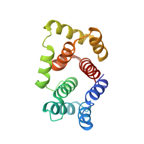
5M1M | pdb_00005m1m

Crystal structure of matrix protein 1 from Influenza C virus (strain C/Ann Arbor/1/1950)


Experimental Data Snapshot

  • Method: X-RAY DIFFRACTION
  • Resolution: 1.50 Å
  • R-Value Free: 
    0.220 (Depositor), 0.215 (DCC) 
  • R-Value Work: 
    0.188 (Depositor), 0.200 (DCC) 
  • R-Value Observed: 
    0.189 (Depositor) 

wwPDB Validation 3D Report Full Report

Validation slider image for 5M1M

This is version 1.2 of the entry. See complete history. 

Literature

The Matrix protein M1 from influenza C virus induces tubular membrane invaginations in an in vitro cell membrane model.

Saletti, D., Radzimanowski, J., Effantin, G., Midtvedt, D., Mangenot, S., Weissenhorn, W., Bassereau, P., Bally, M.

(2017) Sci Rep 7: 40801-40801

  • DOI: https://doi.org/10.1038/srep40801
  • Primary Citation Related Structures: 
    5M1M

  • PubMed Abstract: 

    Matrix proteins from enveloped viruses play an important role in budding and stabilizing virus particles. In order to assess the role of the matrix protein M1 from influenza C virus (M1-C) in plasma membrane deformation, we have combined structural and in vitro reconstitution experiments with model membranes. We present the crystal structure of the N-terminal domain of M1-C and show by Small Angle X-Ray Scattering analysis that full-length M1-C folds into an elongated structure that associates laterally into ring-like or filamentous polymers. Using negatively charged giant unilamellar vesicles (GUVs), we demonstrate that M1-C full-length binds to and induces inward budding of membrane tubules with diameters that resemble the diameter of viruses. Membrane tubule formation requires the C-terminal domain of M1-C, corroborating its essential role for M1-C polymerization. Our results indicate that M1-C assembly on membranes constitutes the driving force for budding and suggest that M1-C plays a key role in facilitating viral egress.


  • Organizational Affiliation: 
    • Laboratoire Physico Chimie Curie, Institut Curie, PSL Research University, CNRS UMR168, 75005, Paris, France.

Macromolecule Content 

  • Total Structure Weight: 17.53 kDa 
  • Atom Count: 1,406 
  • Modeled Residue Count: 154 
  • Deposited Residue Count: 155 
  • Unique protein chains: 1

Macromolecules

Find similar proteins by:|  3D Structure
Entity ID: 1
MoleculeChains  Sequence LengthOrganismDetailsImage
Matrix protein 1155Influenza C virus (C/Ann Arbor/1/50)Mutation(s): 0 
UniProt
Find proteins for Q6I7B9 (Influenza C virus (strain C/Ann Arbor/1/1950))
Explore Q6I7B9 
Go to UniProtKB:  Q6I7B9
Entity Groups
Sequence Clusters30% Identity50% Identity70% Identity90% Identity95% Identity100% Identity
UniProt GroupQ6I7B9
Sequence Annotations
Expand
Reference Sequence

Experimental Data & Validation

Experimental Data

  • Method: X-RAY DIFFRACTION
  • Resolution: 1.50 Å
  • R-Value Free:  0.220 (Depositor), 0.215 (DCC) 
  • R-Value Work:  0.188 (Depositor), 0.200 (DCC) 
  • R-Value Observed: 0.189 (Depositor) 
Space Group: C 1 2 1
Unit Cell:
Length ( Å )Angle ( ˚ )
a = 66.398α = 90
b = 26.841β = 108.4
c = 77.224γ = 90
Software Package:
Software NamePurpose
PHENIXrefinement
XDSdata reduction
SCALAdata scaling
PHENIXphasing

Structure Validation

View Full Validation Report


Entry History 

& Funding Information

Deposition Data


Funding OrganizationLocationGrant Number
French National Research AgencyFrance--

Revision History  (Full details and data files)

  • Version 1.0: 2017-02-08
    Type: Initial release
  • Version 1.1: 2017-09-06
    Changes: Author supporting evidence, Data collection
  • Version 1.2: 2024-05-08
    Changes: Data collection, Database references, Derived calculations