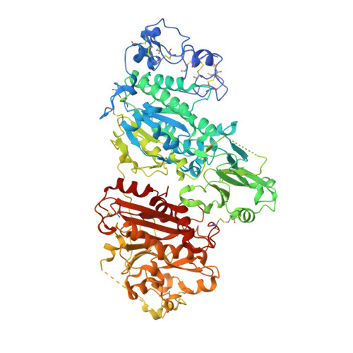
5M0D

Structure-based evolution of a hybrid steroid series of Autotaxin inhibitors


Experimental Data Snapshot

  • Method: X-RAY DIFFRACTION
  • Resolution: 2.40 Å
  • R-Value Free: 0.270 
  • R-Value Work: 0.225 
  • R-Value Observed: 0.227 

Starting Model: experimental
View more details

wwPDB Validation   3D Report Full Report


Ligand Structure Quality Assessment 


This is version 2.1 of the entry. See complete history


Literature

Rational Design of Autotaxin Inhibitors by Structural Evolution of Endogenous Modulators.

Keune, W.J.Potjewyd, F.Heidebrecht, T.Salgado-Polo, F.Macdonald, S.J.Chelvarajan, L.Abdel Latif, A.Soman, S.Morris, A.J.Watson, A.J.Jamieson, C.Perrakis, A.

(2017) J Med Chem 60: 2006-2017

  • DOI: https://doi.org/10.1021/acs.jmedchem.6b01743
  • Primary Citation of Related Structures:  
    5M0D, 5M0E, 5M0M, 5M0S

  • PubMed Abstract: 

    Autotaxin produces the bioactive lipid lysophosphatidic acid (LPA) and is a drug target of considerable interest for numerous pathologies. We report the expedient, structure-guided evolution of weak physiological allosteric inhibitors (bile salts) into potent competitive Autotaxin inhibitors that do not interact with the catalytic site. Functional data confirms that our lead compound attenuates LPA mediated signaling in cells and reduces LPA synthesis in vivo, providing a promising natural product derived scaffold for drug discovery.


  • Organizational Affiliation

    Division of Biochemistry, The Netherlands Cancer Institute , Plesmanlaan 121, 1066CX Amsterdam, The Netherlands.


Macromolecules
Find similar proteins by:  (by identity cutoff)  |  3D Structure
Entity ID: 1
MoleculeChains Sequence LengthOrganismDetailsImage
Ectonucleotide pyrophosphatase/phosphodiesterase family member 2,Ectonucleotide pyrophosphatase/phosphodiesterase family member 2827Rattus norvegicusMutation(s): 3 
Gene Names: Enpp2AtxNpps2
EC: 3.1.4.39 (PDB Primary Data), 3.1.4.4 (UniProt)
UniProt
Find proteins for Q64610 (Rattus norvegicus)
Explore Q64610 
Go to UniProtKB:  Q64610
Entity Groups  
Sequence Clusters30% Identity50% Identity70% Identity90% Identity95% Identity100% Identity
UniProt GroupQ64610
Glycosylation
Glycosylation Sites: 1Go to GlyGen: Q64610-1
Sequence Annotations
Expand
  • Reference Sequence
Oligosaccharides

Help

Entity ID: 2
MoleculeChains Length2D Diagram Glycosylation3D Interactions
alpha-D-mannopyranose-(1-2)-alpha-D-mannopyranose-(1-3)-[alpha-D-mannopyranose-(1-6)]alpha-D-mannopyranose-(1-6)-[alpha-D-mannopyranose-(1-3)]beta-D-mannopyranose-(1-4)-2-acetamido-2-deoxy-beta-D-glucopyranose-(1-4)-2-acetamido-2-deoxy-beta-D-glucopyranose
B
8N-Glycosylation
Glycosylation Resources
GlyTouCan:  G34499SX
GlyCosmos:  G34499SX
GlyGen:  G34499SX
Small Molecules
Ligands 7 Unique
IDChains Name / Formula / InChI Key2D Diagram3D Interactions
7C8
Query on 7C8

Download Ideal Coordinates CCD File 
O [auth A][3,5-bis(chloranyl)phenyl]methyl 4-[(4~{R})-4-[(3~{R},5~{S},7~{S},8~{R},9~{S},10~{S},13~{R},14~{S},17~{R})-10,13-dimethyl-3,7-bis(oxidanyl)-2,3,4,5,6,7,8,9,11,12,14,15,16,17-tetradecahydro-1~{H}-cyclopenta[a]phenanthren-17-yl]pentyl]piperazine-1-carboxylate
C36 H54 Cl2 N2 O4
MXFWEMTZODLDTJ-IPVBVCFFSA-N
IOD
Query on IOD

Download Ideal Coordinates CCD File 
C [auth A]
D [auth A]
E [auth A]
F [auth A]
G [auth A]
C [auth A],
D [auth A],
E [auth A],
F [auth A],
G [auth A],
H [auth A],
I [auth A],
J [auth A],
K [auth A]
IODIDE ION
I
XMBWDFGMSWQBCA-UHFFFAOYSA-M
GOL
Query on GOL

Download Ideal Coordinates CCD File 
U [auth A],
V [auth A],
W [auth A]
GLYCEROL
C3 H8 O3
PEDCQBHIVMGVHV-UHFFFAOYSA-N
ZN
Query on ZN

Download Ideal Coordinates CCD File 
L [auth A],
M [auth A]
ZINC ION
Zn
PTFCDOFLOPIGGS-UHFFFAOYSA-N
SCN
Query on SCN

Download Ideal Coordinates CCD File 
Q [auth A],
R [auth A],
S [auth A],
T [auth A]
THIOCYANATE ION
C N S
ZMZDMBWJUHKJPS-UHFFFAOYSA-M
CA
Query on CA

Download Ideal Coordinates CCD File 
N [auth A]CALCIUM ION
Ca
BHPQYMZQTOCNFJ-UHFFFAOYSA-N
NA
Query on NA

Download Ideal Coordinates CCD File 
P [auth A]SODIUM ION
Na
FKNQFGJONOIPTF-UHFFFAOYSA-N
Experimental Data & Validation

Experimental Data

  • Method: X-RAY DIFFRACTION
  • Resolution: 2.40 Å
  • R-Value Free: 0.270 
  • R-Value Work: 0.225 
  • R-Value Observed: 0.227 
  • Space Group: P 1 21 1
Unit Cell:
Length ( Å )Angle ( ˚ )
a = 62.927α = 90
b = 88.012β = 103.1
c = 77.356γ = 90
Software Package:
Software NamePurpose
REFMACrefinement
XDSdata reduction
Aimlessdata scaling
PHASERphasing

Structure Validation

View Full Validation Report


Ligand Structure Quality Assessment 


Entry History 

Deposition Data

Revision History  (Full details and data files)

  • Version 1.0: 2017-08-16
    Type: Initial release
  • Version 1.1: 2019-10-16
    Changes: Data collection
  • Version 2.0: 2020-07-29
    Type: Remediation
    Reason: Carbohydrate remediation
    Changes: Atomic model, Data collection, Derived calculations, Structure summary
  • Version 2.1: 2024-01-17
    Changes: Data collection, Database references, Derived calculations, Refinement description, Structure summary