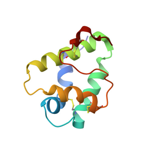
Ligand Binding Properties of the Lentil Lipid Transfer Protein: Molecular Insight into the Possible Mechanism of Lipid Uptake.
Shenkarev, Z.O., Melnikova, D.N., Finkina, E.I., Sukhanov, S.V., Boldyrev, I.A., Gizatullina, A.K., Mineev, K.S., Arseniev, A.S., Ovchinnikova, T.V.(2017) Biochemistry 56: 1785-1796
- PubMed: 28266846
- DOI: https://doi.org/10.1021/acs.biochem.6b01079
- Primary Citation of Related Structures:
5LQV - PubMed Abstract:
The lentil lipid transfer protein, designated as Lc-LTP2, was isolated from Lens culinaris seeds. The protein belongs to the LTP1 subfamily and consists of 93 amino acid residues. Its spatial structure includes four α-helices (H1-H4) and a long C-terminal tail. Here, we report the ligand binding properties of Lc-LTP2. The fluorescent 2-p-toluidinonaphthalene-6-sulfonate binding assay revealed that the affinity of Lc-LTP2 for saturated and unsaturated fatty acids was enhanced with a decrease in acyl-chain length. Measurements of boundary potential in planar lipid bilayers and calcein dye leakage in vesicular systems revealed preferential interaction of Lc-LTP2 with the negatively charged membranes. Lc-LTP2 more efficiently transferred anionic dimyristoylphosphatidylglycerol (DMPG) than zwitterionic dimyristoylphosphatidylcholine. Nuclear magnetic resonance experiments confirmed the higher affinity of Lc-LTP2 for anionic lipids and those with smaller volumes of hydrophobic chains. The acyl chains of the bound lysopalmitoylphosphatidylglycerol (LPPG), DMPG, or dihexanoylphosphatidylcholine molecules occupied the internal hydrophobic cavity, while their headgroups protruded into the aqueous environment between helices H1 and H3. The spatial structure and backbone dynamics of the Lc-LTP2-LPPG complex were determined. The internal cavity was expanded from ∼600 to ∼1000 Å 3 upon the ligand binding. Another entrance into the internal cavity, restricted by the H2-H3 interhelical loop and C-terminal tail, appeared to be responsible for the attachment of Lc-LTP2 to the membrane or micelle surface and probably played an important role in the lipid uptake determining the ligand specificity. Our results confirmed the previous assumption regarding the membrane-mediated antimicrobial action of Lc-LTP2 and afforded molecular insight into its biological role in the plant.
- M. M. Shemyakin and Yu. A. Ovchinnikov Institute of Bioorganic Chemistry, Russian Academy of Sciences , Miklukho-Maklaya street, 16/10, 117997 Moscow, Russia.
Organizational Affiliation:


