5LQQ | pdb_00005lqq

Structure of Autotaxin (ENPP2) with LM350


Experimental Data Snapshot

  • Method: X-RAY DIFFRACTION
  • Resolution: 2.40 Å
  • R-Value Free: 
    0.262 (Depositor), 0.227 (DCC) 
  • R-Value Work: 
    0.217 (Depositor) 
  • R-Value Observed: 
    0.219 (Depositor) 

Starting Model: experimental
View more details

wwPDB Validation 3D Report Full Report

Validation slider image for 5LQQ

Ligand Structure Quality Assessment 


This is version 2.2 of the entry. See complete history

Literature

Structure-Activity Relationships of Small Molecule Autotaxin Inhibitors with a Discrete Binding Mode.

Miller, L.M.Keune, W.J.Castagna, D.Young, L.C.Duffy, E.L.Potjewyd, F.Salgado-Polo, F.Engel Garcia, P.Semaan, D.Pritchard, J.M.Perrakis, A.Macdonald, S.J.Jamieson, C.Watson, A.J.

(2017) J Med Chem 60: 722-748

  • DOI: https://doi.org/10.1021/acs.jmedchem.6b01597
  • Primary Citation Related Structures: 
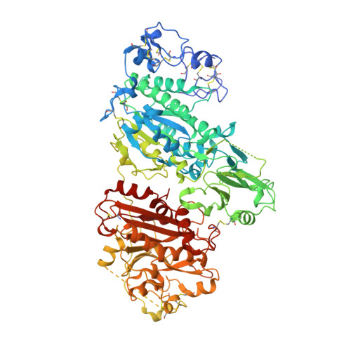
    5LQQ

  • PubMed Abstract: 

    Autotaxin (ATX) is a secreted enzyme responsible for the hydrolysis of lysophosphatidylcholine (LPC) to the bioactive lysophosphatidic acid (LPA) and choline. The ATX-LPA signaling pathway is implicated in cell survival, migration, and proliferation; thus, the inhibition of ATX is a recognized therapeutic target for a number of diseases including fibrotic diseases, cancer, and inflammation, among others. Many of the developed synthetic inhibitors for ATX have resembled the lipid chemotype of the native ligand; however, a small number of inhibitors have been described that deviate from this common scaffold. Herein, we report the structure-activity relationships (SAR) of a previously reported small molecule ATX inhibitor. We show through enzyme kinetics studies that analogues of this chemotype are noncompetitive inhibitors, and by using a crystal structure with ATX we confirm the discrete binding mode.


  • Organizational Affiliation
    • WestCHEM, Department of Pure and Applied Chemistry, University of Strathclyde , Thomas Graham Building, 295 Cathedral Street, Glasgow G1 1XL, U.K.

Macromolecule Content 

  • Total Structure Weight: 98.29 kDa 
  • Atom Count: 6,466 
  • Modeled Residue Count: 775 
  • Deposited Residue Count: 827 
  • Unique protein chains: 1

Macromolecules

Find similar proteins by:|  3D Structure
Entity ID: 1
MoleculeChains  Sequence LengthOrganismDetailsImage
Ectonucleotide pyrophosphatase/phosphodiesterase family member 2827Rattus norvegicusMutation(s): 2 
Gene Names: Enpp2AtxNpps2
EC: 3.1.4.39 (PDB Primary Data), 3.1.4.4 (UniProt)
UniProt
Find proteins for Q64610 (Rattus norvegicus)
Explore Q64610 
Go to UniProtKB:  Q64610
Entity Groups
Sequence Clusters30% Identity50% Identity70% Identity90% Identity95% Identity100% Identity
UniProt GroupQ64610
Glycosylation
Glycosylation Sites: 1Go to GlyGen: Q64610-1
Sequence Annotations
Expand
Reference Sequence

Oligosaccharides

Help  
Entity ID: 2
MoleculeChains Length2D Diagram GlycosylationD Interactions
alpha-D-mannopyranose-(1-2)-alpha-D-mannopyranose-(1-3)-[alpha-D-mannopyranose-(1-6)]alpha-D-mannopyranose-(1-6)-[alpha-D-mannopyranose-(1-3)]beta-D-mannopyranose-(1-4)-2-acetamido-2-deoxy-beta-D-glucopyranose-(1-4)-2-acetamido-2-deoxy-beta-D-glucopyranose
B
8N-Glycosylation
Glycosylation Resources
GlyTouCan: G34499SX
GlyCosmos: G34499SX
GlyGen: G34499SX

Small Molecules

Ligands 5 Unique
IDChains Name / Formula / InChI Key2D Diagram3D Interactions
72P

Query on 72P


Download:Ideal Coordinates CCD File
P [auth A]3-(6-chloranyl-2-methyl-1-phenyl-indol-3-yl)sulfanylbenzoic acid
C22 H16 Cl N O2 S
QESQUQBNRUZICQ-UHFFFAOYSA-N
IOD

Query on IOD


Download:Ideal Coordinates CCD File
F [auth A]
G [auth A]
H [auth A]
I [auth A]
J [auth A]
F [auth A],
G [auth A],
H [auth A],
I [auth A],
J [auth A],
K [auth A],
L [auth A],
M [auth A],
N [auth A],
O [auth A]
IODIDE ION
I
XMBWDFGMSWQBCA-UHFFFAOYSA-M
ZN

Query on ZN


Download:Ideal Coordinates CCD File
C [auth A],
D [auth A]
ZINC ION
Zn
PTFCDOFLOPIGGS-UHFFFAOYSA-N
CA

Query on CA


Download:Ideal Coordinates CCD File
E [auth A]CALCIUM ION
Ca
BHPQYMZQTOCNFJ-UHFFFAOYSA-N
NA

Query on NA


Download:Ideal Coordinates CCD File
Q [auth A]SODIUM ION
Na
FKNQFGJONOIPTF-UHFFFAOYSA-N
Binding Affinity Annotations 
IDSourceBinding Affinity
72P BindingDB:  5LQQ IC50: min: 5, max: 300 (nM) from 3 assay(s)

Experimental Data & Validation

Experimental Data

  • Method: X-RAY DIFFRACTION
  • Resolution: 2.40 Å
  • R-Value Free:  0.262 (Depositor), 0.227 (DCC) 
  • R-Value Work:  0.217 (Depositor) 
  • R-Value Observed: 0.219 (Depositor) 
Space Group: P 1 21 1
Unit Cell:
Length ( Å )Angle ( ˚ )
a = 62.655α = 90
b = 89.067β = 102.88
c = 77.509γ = 90
Software Package:
Software NamePurpose
Aimlessdata scaling
PHASERphasing
REFMACrefinement
PDB_EXTRACTdata extraction
XDSdata reduction

Structure Validation

View Full Validation Report


Ligand Structure Quality Assessment 


Entry History 

Deposition Data

Revision History  (Full details and data files)

  • Version 1.0: 2016-12-28
    Type: Initial release
  • Version 1.1: 2017-01-18
    Changes: Database references
  • Version 1.2: 2017-02-08
    Changes: Database references
  • Version 2.0: 2020-07-29
    Type: Remediation
    Reason: Carbohydrate remediation
    Changes: Advisory, Atomic model, Data collection, Derived calculations, Structure summary
  • Version 2.1: 2024-01-10
    Changes: Data collection, Database references, Derived calculations, Refinement description, Structure summary
  • Version 2.2: 2024-10-16
    Changes: Structure summary