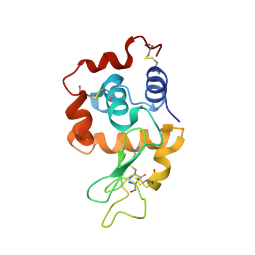
5LIO

Lysozyme, collected at rotation 360 degree per second


Experimental Data Snapshot

  • Method: X-RAY DIFFRACTION
  • Resolution: 1.50 Å
  • R-Value Free: 0.249 
  • R-Value Work: 0.195 
  • R-Value Observed: 0.197 

Starting Model: experimental
View more details

wwPDB Validation   3D Report Full Report


This is version 1.4 of the entry. See complete history


Literature

EIGER detector: application in macromolecular crystallography.

Casanas, A.Warshamanage, R.Finke, A.D.Panepucci, E.Olieric, V.Noll, A.Tampe, R.Brandstetter, S.Forster, A.Mueller, M.Schulze-Briese, C.Bunk, O.Wang, M.

(2016) Acta Crystallogr D Struct Biol 72: 1036-1048

  • DOI: https://doi.org/10.1107/S2059798316012304
  • Primary Citation of Related Structures:  
    5LIN, 5LIO, 5LIS

  • PubMed Abstract: 

    The development of single-photon-counting detectors, such as the PILATUS, has been a major recent breakthrough in macromolecular crystallography, enabling noise-free detection and novel data-acquisition modes. The new EIGER detector features a pixel size of 75 × 75 µm, frame rates of up to 3000 Hz and a dead time as low as 3.8 µs. An EIGER 1M and EIGER 16M were tested on Swiss Light Source beamlines X10SA and X06SA for their application in macromolecular crystallography. The combination of fast frame rates and a very short dead time allows high-quality data acquisition in a shorter time. The ultrafine ϕ-slicing data-collection method is introduced and validated and its application in finding the optimal rotation angle, a suitable rotation speed and a sufficient X-ray dose are presented. An improvement of the data quality up to slicing at one tenth of the mosaicity has been observed, which is much finer than expected based on previous findings. The influence of key data-collection parameters on data quality is discussed.


  • Organizational Affiliation

    Swiss Light Source, Paul Scherrer Institute, 5232 Villigen, Switzerland.


Macromolecules
Find similar proteins by:  (by identity cutoff)  |  3D Structure
Entity ID: 1
MoleculeChains Sequence LengthOrganismDetailsImage
Lysozyme C129Gallus gallusMutation(s): 0 
EC: 3.2.1.17
UniProt
Find proteins for P00698 (Gallus gallus)
Explore P00698 
Go to UniProtKB:  P00698
Entity Groups  
Sequence Clusters30% Identity50% Identity70% Identity90% Identity95% Identity100% Identity
UniProt GroupP00698
Sequence Annotations
Expand
  • Reference Sequence
Experimental Data & Validation

Experimental Data

  • Method: X-RAY DIFFRACTION
  • Resolution: 1.50 Å
  • R-Value Free: 0.249 
  • R-Value Work: 0.195 
  • R-Value Observed: 0.197 
  • Space Group: P 43 21 2
Unit Cell:
Length ( Å )Angle ( ˚ )
a = 78.37α = 90
b = 78.37β = 90
c = 37.044γ = 90
Software Package:
Software NamePurpose
REFMACrefinement
XDSdata reduction
XDSdata scaling
MOLREPphasing

Structure Validation

View Full Validation Report


Entry History 

Deposition Data

Revision History  (Full details and data files)

  • Version 1.0: 2016-08-03
    Type: Initial release
  • Version 1.1: 2016-09-14
    Changes: Database references
  • Version 1.2: 2016-09-28
    Changes: Database references
  • Version 1.3: 2024-01-10
    Changes: Data collection, Database references, Refinement description
  • Version 1.4: 2024-11-06
    Changes: Structure summary