5LB1 | pdb_00005lb1

Crystal structure of the Mycobacterium tuberculosis L,D-transpeptidase-2 (LdtMt2) BC-module with thionitrobenzoate (TNB) adduct at the active site cysteine-354


Experimental Data Snapshot

  • Method: X-RAY DIFFRACTION
  • Resolution: 1.55 Å
  • R-Value Free: 
    0.194 (Depositor), 0.203 (DCC) 
  • R-Value Work: 
    0.147 (Depositor), 0.160 (DCC) 
  • R-Value Observed: 
    0.150 (Depositor) 

Starting Model: experimental
View more details

wwPDB Validation 3D Report Full Report

Validation slider image for 5LB1

Ligand Structure Quality Assessment 


This is version 1.3 of the entry. See complete history

Literature

Binding and processing of beta-lactam antibiotics by the transpeptidase LdtMt2 from Mycobacterium tuberculosis.

Steiner, E.M.Schneider, G.Schnell, R.

(2017) FEBS J 284: 725-741

  • DOI: https://doi.org/10.1111/febs.14010
  • Primary Citation Related Structures: 
    5LB1, 5LBG

  • PubMed Abstract: 

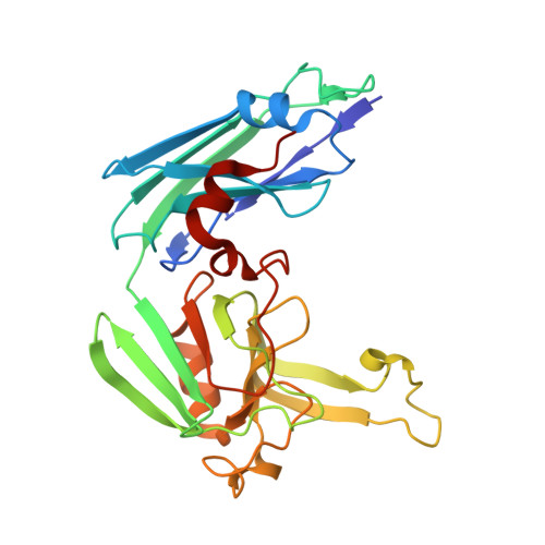
    β-lactam antibiotics represent a novel direction in the chemotherapy of tuberculosis that brings the peptidoglycan layer of the complex mycobacterial cell wall in focus as a therapeutic target. Peptidoglycan stability in Mycobacterium tuberculosis, especially during infection, relies on the nonconventional peptide cross-links formed by l,d-transpeptidases. These enzymes are known to be inhibited by β-lactams, primarily carbapenems, leading to a stable covalent modification at the enzyme active site. A panel of 16 β-lactam antibiotics was characterized by inhibition kinetics, mass spectrometry, and x-ray crystallography to identify efficient compounds and study their action on the essential transpeptidase, Ldt Mt2 . Members of the carbapenem class displayed fast binding kinetics, but faropenem, a penem type compound showed a three to four time higher rate in the adduct formation. In three cases, mass spectrometry indicated that carbapenems may undergo decarboxylation, while faropenem decomposition following the acylation step results in a small 87 Da β-OH-butyryl adduct bound at the catalytic cysteine residue. The crystal structure of Ldt Mt2 at 1.54 Å resolution with this fragment bound revealed that the protein adopts a closed conformation that shields the thioester bond from the solvent, which is in line with the high stability of this dead-end complex observed also in biochemical assays. Structural data are available in Protein Data Bank under the accession numbers 5LB1 and 5LBG.


  • Organizational Affiliation
    • Department of Medical Biochemistry and Biophysics, Karolinska Institutet, Stockholm, Sweden.

Macromolecule Content 

  • Total Structure Weight: 57.42 kDa 
  • Atom Count: 4,789 
  • Modeled Residue Count: 511 
  • Deposited Residue Count: 524 
  • Unique protein chains: 1

Macromolecules

Find similar proteins by:|  3D Structure
Entity ID: 1
MoleculeChains  Sequence LengthOrganismDetailsImage
L,D-transpeptidase 2
A, B
262Mycobacterium tuberculosis H37RvMutation(s): 0 
Gene Names: ldtBlppSRv2518cRVBD_2518cP425_02624
EC: 2.3.2
UniProt
Find proteins for I6Y9J2 (Mycobacterium tuberculosis (strain ATCC 25618 / H37Rv))
Explore I6Y9J2 
Go to UniProtKB:  I6Y9J2
Entity Groups
Sequence Clusters30% Identity50% Identity70% Identity90% Identity95% Identity100% Identity
UniProt GroupI6Y9J2
Sequence Annotations
Expand
Reference Sequence

Experimental Data & Validation

Experimental Data

  • Method: X-RAY DIFFRACTION
  • Resolution: 1.55 Å
  • R-Value Free:  0.194 (Depositor), 0.203 (DCC) 
  • R-Value Work:  0.147 (Depositor), 0.160 (DCC) 
  • R-Value Observed: 0.150 (Depositor) 
Space Group: P 1
Unit Cell:
Length ( Å )Angle ( ˚ )
a = 53.368α = 68.84
b = 53.91β = 75.03
c = 57.174γ = 88.64
Software Package:
Software NamePurpose
REFMACrefinement
XDSdata reduction
Aimlessdata scaling
MOLREPphasing

Structure Validation

View Full Validation Report


Ligand Structure Quality Assessment 


Entry History 

& Funding Information

Deposition Data


Funding OrganizationLocationGrant Number
Research CouncilSweden--

Revision History  (Full details and data files)

  • Version 1.0: 2017-01-18
    Type: Initial release
  • Version 1.1: 2017-03-15
    Changes: Database references
  • Version 1.2: 2024-01-10
    Changes: Data collection, Database references, Refinement description
  • Version 1.3: 2024-10-23
    Changes: Structure summary