5L86 | pdb_00005l86

engineered ascorbate peroxidise


Experimental Data Snapshot

  • Method: X-RAY DIFFRACTION
  • Resolution: 1.90 Å
  • R-Value Free: 
    0.231 (Depositor), 0.231 (DCC) 
  • R-Value Work: 
    0.179 (Depositor), 0.179 (DCC) 
  • R-Value Observed: 
    0.182 (Depositor) 

Starting Model: experimental
View more details

wwPDB Validation 3D Report Full Report

Validation slider image for 5L86

Ligand Structure Quality Assessment 


This is version 1.2 of the entry. See complete history

Literature

A Chemically Programmed Proximal Ligand Enhances the Catalytic Properties of a Heme Enzyme.

Green, A.P.Hayashi, T.Mittl, P.R.Hilvert, D.

(2016) J Am Chem Soc 138: 11344-11352

  • DOI: https://doi.org/10.1021/jacs.6b07029
  • Primary Citation Related Structures: 
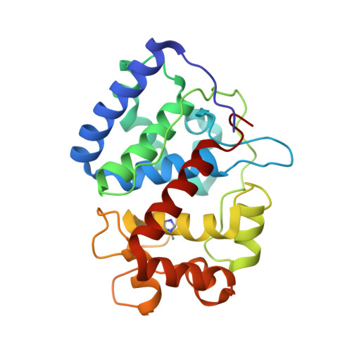
    5L86

  • PubMed Abstract: 

    Enzymes rely on complex interactions between precisely positioned active site residues as a mechanism to compensate for the limited functionality contained within the genetic code. Heme enzymes provide a striking example of this complexity, whereby the electronic properties of reactive ferryl intermediates are finely tuned through hydrogen bonding interactions between proximal ligands and neighboring amino acids. Here, we show that introduction of a chemically programmed proximal Nδ-methyl histidine (NMH) ligand into an engineered ascorbate peroxidase (APX2) overcomes the reliance on the conserved Asp-His hydrogen bonding interaction, leading to a catalytically modified enzyme (APX2 NMH), which is able to achieve a significantly higher number of turnovers compared with APX2 without compromising catalytic efficiency. Structural, spectroscopic and kinetic characterization of APX2 NMH and several active site variants provides valuable insights into the role of the Asp-His-Fe triad of heme peroxidases. More significantly, simplification of catalytic mechanisms through the incorporation of chemically optimized ligands may facilitate efforts to create and evolve new active site heme environments within proteins.


  • Organizational Affiliation
    • School of Chemistry & Manchester Institute of Biotechnology, The University of Manchester , 131 Princess Street, Manchester M1 7DN, U.K.

Macromolecule Content 

  • Total Structure Weight: 55.44 kDa 
  • Atom Count: 4,289 
  • Modeled Residue Count: 494 
  • Deposited Residue Count: 494 
  • Unique protein chains: 1

Macromolecules

Find similar proteins by:|  3D Structure
Entity ID: 1
MoleculeChains  Sequence LengthOrganismDetailsImage
Ascorbate peroxidase
A, B
247Glycine maxMutation(s): 4 
Gene Names: apx1GLYMA_U021900
EC: 1.11.1.11
UniProt
Find proteins for Q43758 (Glycine max)
Explore Q43758 
Go to UniProtKB:  Q43758
Entity Groups
Sequence Clusters30% Identity50% Identity70% Identity90% Identity95% Identity100% Identity
UniProt GroupQ43758
Sequence Annotations
Expand
Reference Sequence

Small Molecules

Ligands 2 Unique
IDChains Name / Formula / InChI Key2D Diagram3D Interactions
HEM

Query on HEM


Download:Ideal Coordinates CCD File
C [auth A],
G [auth B]
PROTOPORPHYRIN IX CONTAINING FE
C34 H32 Fe N4 O4
KABFMIBPWCXCRK-RGGAHWMASA-L
SO4

Query on SO4


Download:Ideal Coordinates CCD File
D [auth A]
E [auth A]
F [auth A]
H [auth B]
I [auth B]
D [auth A],
E [auth A],
F [auth A],
H [auth B],
I [auth B],
J [auth B]
SULFATE ION
O4 S
QAOWNCQODCNURD-UHFFFAOYSA-L
Modified Residues  1 Unique
IDChains TypeFormula2D DiagramParent
MHS
Query on MHS
A, B
L-PEPTIDE LINKINGC7 H11 N3 O2HIS

Experimental Data & Validation

Experimental Data

  • Method: X-RAY DIFFRACTION
  • Resolution: 1.90 Å
  • R-Value Free:  0.231 (Depositor), 0.231 (DCC) 
  • R-Value Work:  0.179 (Depositor), 0.179 (DCC) 
  • R-Value Observed: 0.182 (Depositor) 
Space Group: P 21 21 21
Unit Cell:
Length ( Å )Angle ( ˚ )
a = 84.46α = 90
b = 81.95β = 90
c = 69.8γ = 90
Software Package:
Software NamePurpose
XSCALEdata scaling
REFMACrefinement
PDB_EXTRACTdata extraction
XDSdata reduction
MOLREPphasing

Structure Validation

View Full Validation Report


Ligand Structure Quality Assessment 


Entry History 

Deposition Data

Revision History  (Full details and data files)

  • Version 1.0: 2017-03-01
    Type: Initial release
  • Version 1.1: 2017-03-08
    Changes: Database references
  • Version 1.2: 2024-01-10
    Changes: Data collection, Database references, Derived calculations, Refinement description