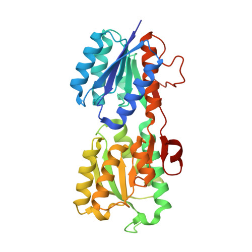
Crystal Structure of Galactose Binding Protein from Yersinia pestis in the Complex with beta D Glucose
Kim, Y., Maltseva, N., Mulligan, R., Grimshaw, S., Anderson, W.F., Joachimiak, A., Center for Structural Genomics of Infectious Diseases (CSGID)To be published.