


Structure of Malate Dehydrogenase in complex with NADH from Mycobacterium tuberculosis
Cheng, Y.S., Sacchettini, J.C.To be published.
Experimental Data Snapshot
Starting Model: experimental
View more details
Entity ID: 1 | |||||
|---|---|---|---|---|---|
| Molecule | Chains | Sequence Length | Organism | Details | Image |
| Malate dehydrogenase | 335 | Mycobacterium tuberculosis H37Rv | Mutation(s): 0 Gene Names: mdh, Rv1240, MTV006.12 EC: 1.1.1.37 | 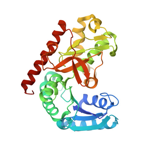 | |
UniProt | |||||
Entity Groups | |||||
| Sequence Clusters | 30% Identity50% Identity70% Identity90% Identity95% Identity100% Identity | ||||
| UniProt Group | P9WK13 | ||||
Sequence AnnotationsExpand | |||||
Reference Sequence | |||||
| Ligands 4 Unique | |||||
|---|---|---|---|---|---|
| ID | Chains | Name / Formula / InChI Key | 2D Diagram | 3D Interactions | |
| NAI Download:Ideal Coordinates CCD File | C [auth A], F [auth B] | 1,4-DIHYDRONICOTINAMIDE ADENINE DINUCLEOTIDE C21 H29 N7 O14 P2 BOPGDPNILDQYTO-NNYOXOHSSA-N |  | ||
| TRS Download:Ideal Coordinates CCD File | E [auth A] | 2-AMINO-2-HYDROXYMETHYL-PROPANE-1,3-DIOL C4 H12 N O3 LENZDBCJOHFCAS-UHFFFAOYSA-O |  | ||
| SO4 Download:Ideal Coordinates CCD File | H [auth B], I [auth B] | SULFATE ION O4 S QAOWNCQODCNURD-UHFFFAOYSA-L |  | ||
| GOL Download:Ideal Coordinates CCD File | D [auth A], G [auth B] | GLYCEROL C3 H8 O3 PEDCQBHIVMGVHV-UHFFFAOYSA-N |  | ||
| Length ( Å ) | Angle ( ˚ ) |
|---|---|
| a = 52.206 | α = 90 |
| b = 78.169 | β = 90 |
| c = 154.402 | γ = 90 |
| Software Name | Purpose |
|---|---|
| HKL-2000 | data scaling |
| PHENIX | refinement |
| PDB_EXTRACT | data extraction |
| HKL-2000 | data reduction |
| MOLREP | phasing |


