


Structural and kinetic characterization of isocitrate dehydrogenase-2 from Mycobacterium tuberculosis
Sacchettini, J.C., Cheng, Y.S.To be published.
Experimental Data Snapshot
Starting Model: experimental
View more details
Entity ID: 1 | |||||
|---|---|---|---|---|---|
| Molecule | Chains | Sequence Length | Organism | Details | Image |
| Isocitrate dehydrogenase | 745 | Mycobacterium tuberculosis H37Rv | Mutation(s): 0 Gene Names: icd2, Rv0066c, LH57_00380 EC: 1.1.1.42 | 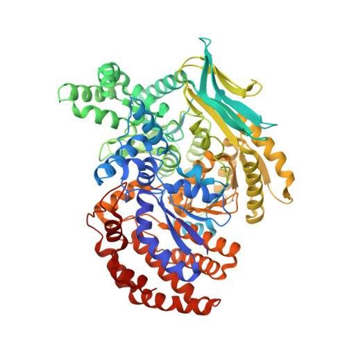 | |
UniProt | |||||
Entity Groups | |||||
| Sequence Clusters | 30% Identity50% Identity70% Identity90% Identity95% Identity100% Identity | ||||
| UniProt Group | O53611 | ||||
Sequence AnnotationsExpand | |||||
Reference Sequence | |||||
| Ligands 6 Unique | |||||
|---|---|---|---|---|---|
| ID | Chains | Name / Formula / InChI Key | 2D Diagram | 3D Interactions | |
| NAP Download:Ideal Coordinates CCD File | E [auth A], H [auth B], L [auth C], Q [auth D] | NADP NICOTINAMIDE-ADENINE-DINUCLEOTIDE PHOSPHATE C21 H28 N7 O17 P3 XJLXINKUBYWONI-NNYOXOHSSA-N |  | ||
| MLT Download:Ideal Coordinates CCD File | F [auth A] | D-MALATE C4 H6 O5 BJEPYKJPYRNKOW-UWTATZPHSA-N |  | ||
| SIN Download:Ideal Coordinates CCD File | M [auth C] | SUCCINIC ACID C4 H6 O4 KDYFGRWQOYBRFD-UHFFFAOYSA-N |  | ||
| MLA Download:Ideal Coordinates CCD File | I [auth B], R [auth D] | MALONIC ACID C3 H4 O4 OFOBLEOULBTSOW-UHFFFAOYSA-N |  | ||
| GOL Download:Ideal Coordinates CCD File | N [auth C], S [auth D] | GLYCEROL C3 H8 O3 PEDCQBHIVMGVHV-UHFFFAOYSA-N |  | ||
| EDO Download:Ideal Coordinates CCD File | G [auth A] J [auth B] K [auth B] O [auth C] P [auth C] | 1,2-ETHANEDIOL C2 H6 O2 LYCAIKOWRPUZTN-UHFFFAOYSA-N |  | ||
| Length ( Å ) | Angle ( ˚ ) |
|---|---|
| a = 74.827 | α = 86.01 |
| b = 110.861 | β = 78.62 |
| c = 114.914 | γ = 76.79 |
| Software Name | Purpose |
|---|---|
| PHENIX | refinement |
| HKL-3000 | data collection |
| HKL-2000 | data scaling |
| PDB_EXTRACT | data extraction |
| HKL-3000 | data reduction |
| MOLREP | phasing |


