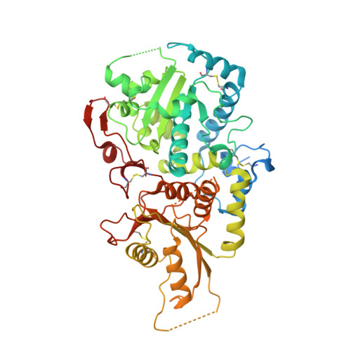
5KOR

Arabidopsis thaliana fucosyltransferase 1 (FUT1) in complex with GDP and a xylo-oligossacharide


Experimental Data Snapshot

  • Method: X-RAY DIFFRACTION
  • Resolution: 2.20 Å
  • R-Value Free: 0.215 
  • R-Value Work: 0.179 
  • R-Value Observed: 0.181 

Starting Model: experimental
View more details

wwPDB Validation   3D Report Full Report


Ligand Structure Quality Assessment 


This is version 2.1 of the entry. See complete history


Literature

Structure of Arabidopsis thaliana FUT1 Reveals a Variant of the GT-B Class Fold and Provides Insight into Xyloglucan Fucosylation.

Rocha, J.Ciceron, F.de Sanctis, D.Lelimousin, M.Chazalet, V.Lerouxel, O.Breton, C.

(2016) Plant Cell 28: 2352-2364

  • DOI: https://doi.org/10.1105/tpc.16.00519
  • Primary Citation of Related Structures:  
    5KOP, 5KOR

  • PubMed Abstract: 

    The plant cell wall is a complex and dynamic network made mostly of cellulose, hemicelluloses, and pectins. Xyloglucan, the major hemicellulosic component in Arabidopsis thaliana, is biosynthesized in the Golgi apparatus by a series of glycan synthases and glycosyltransferases before export to the wall. A better understanding of the xyloglucan biosynthetic machinery will give clues toward engineering plants with improved wall properties or designing novel xyloglucan-based biomaterials. The xyloglucan-specific α2-fucosyltransferase FUT1 catalyzes the transfer of fucose from GDP-fucose to terminal galactosyl residues on xyloglucan side chains. Here, we present crystal structures of Arabidopsis FUT1 in its apoform and in a ternary complex with GDP and a xylo-oligosaccharide acceptor (named XLLG). Although FUT1 is clearly a member of the large GT-B fold family, like other fucosyltransferases of known structures, it contains a variant of the GT-B fold. In particular, it includes an extra C-terminal region that is part of the acceptor binding site. Our crystal structures support previous findings that FUT1 behaves as a functional dimer. Mutational studies and structure comparison with other fucosyltransferases suggest that FUT1 uses a S N 2-like reaction mechanism similar to that of protein-O-fucosyltransferase 2. Thus, our results provide new insights into the mechanism of xyloglucan fucosylation in the Golgi.


  • Organizational Affiliation

    Université Grenoble Alpes, 38400 Grenoble, France.


Macromolecules
Find similar proteins by:  (by identity cutoff)  |  3D Structure
Entity ID: 1
MoleculeChains Sequence LengthOrganismDetailsImage
Galactoside 2-alpha-L-fucosyltransferase
A, B, C, D
521Arabidopsis thalianaMutation(s): 0 
Gene Names: FUT1FT1MUR2At2g03220T18E12.11
EC: 2.4.1.69
UniProt
Find proteins for Q9SWH5 (Arabidopsis thaliana)
Explore Q9SWH5 
Go to UniProtKB:  Q9SWH5
Entity Groups  
Sequence Clusters30% Identity50% Identity70% Identity90% Identity95% Identity100% Identity
UniProt GroupQ9SWH5
Sequence Annotations
Expand
  • Reference Sequence
Oligosaccharides

Help

Entity ID: 2
MoleculeChains Length2D Diagram Glycosylation3D Interactions
alpha-D-xylopyranose-(1-6)-beta-D-glucopyranose-(1-4)-[beta-D-galactopyranose-(1-2)-alpha-D-xylopyranose-(1-6)]beta-D-glucopyranose-(1-4)-[beta-D-galactopyranose-(1-2)-alpha-D-xylopyranose-(1-6)]beta-D-glucopyranose-(1-4)-beta-D-glucopyranose
E, F
9N/A
Glycosylation Resources
GlyTouCan:  G06184LI
GlyCosmos:  G06184LI
Small Molecules
Ligands 3 Unique
IDChains Name / Formula / InChI Key2D Diagram3D Interactions
GDP
Query on GDP

Download Ideal Coordinates CCD File 
G [auth A],
K [auth B],
O [auth C],
W [auth D]
GUANOSINE-5'-DIPHOSPHATE
C10 H15 N5 O11 P2
QGWNDRXFNXRZMB-UUOKFMHZSA-N
EDO
Query on EDO

Download Ideal Coordinates CCD File 
H [auth A]
I [auth A]
J [auth A]
L [auth B]
M [auth B]
H [auth A],
I [auth A],
J [auth A],
L [auth B],
M [auth B],
P [auth C],
Q [auth C],
R [auth C],
S [auth C],
T [auth C],
U [auth C],
X [auth D],
Y [auth D],
Z [auth D]
1,2-ETHANEDIOL
C2 H6 O2
LYCAIKOWRPUZTN-UHFFFAOYSA-N
CL
Query on CL

Download Ideal Coordinates CCD File 
AA [auth D],
N [auth B],
V [auth C]
CHLORIDE ION
Cl
VEXZGXHMUGYJMC-UHFFFAOYSA-M
Experimental Data & Validation

Experimental Data

  • Method: X-RAY DIFFRACTION
  • Resolution: 2.20 Å
  • R-Value Free: 0.215 
  • R-Value Work: 0.179 
  • R-Value Observed: 0.181 
  • Space Group: P 1 21 1
Unit Cell:
Length ( Å )Angle ( ˚ )
a = 87.975α = 90
b = 85.806β = 96.19
c = 150.723γ = 90
Software Package:
Software NamePurpose
PHENIXrefinement
XDSdata reduction
Aimlessdata scaling
PHASERphasing

Structure Validation

View Full Validation Report


Ligand Structure Quality Assessment 


Entry History & Funding Information

Deposition Data


Funding OrganizationLocationGrant Number
Univ. Grenoble AlpesFrance--

Revision History  (Full details and data files)

  • Version 1.0: 2016-10-12
    Type: Initial release
  • Version 1.1: 2016-11-23
    Changes: Database references
  • Version 1.2: 2018-10-10
    Changes: Data collection, Database references, Source and taxonomy, Structure summary
  • Version 2.0: 2020-07-29
    Type: Remediation
    Reason: Carbohydrate remediation
    Changes: Atomic model, Data collection, Derived calculations, Structure summary
  • Version 2.1: 2024-01-10
    Changes: Data collection, Database references, Derived calculations, Refinement description, Structure summary