


Crystal structure of Mycobacterium tuberculosis hypoxanthine guanine phosphoribosyltransferase in complex with pyrrolidine nucleoside phosphonate
Eng, W.S., Rejman, D., Keough, D.T., Guddat, L.W.To be published.
Experimental Data Snapshot
Entity ID: 1 | |||||
|---|---|---|---|---|---|
| Molecule | Chains | Sequence Length | Organism | Details | Image |
| Hypoxanthine-guanine phosphoribosyltransferase | 207 | Mycobacterium tuberculosis H37Rv | Mutation(s): 0 Gene Names: hpt, hprT, Rv3624c, MTCY15C10.28 EC: 2.4.2.8 | 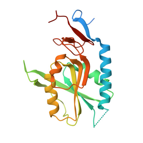 | |
UniProt | |||||
Entity Groups | |||||
| Sequence Clusters | 30% Identity50% Identity70% Identity90% Identity95% Identity100% Identity | ||||
| UniProt Group | P9WHQ9 | ||||
Sequence AnnotationsExpand | |||||
Reference Sequence | |||||
| Ligands 2 Unique | |||||
|---|---|---|---|---|---|
| ID | Chains | Name / Formula / InChI Key | 2D Diagram | 3D Interactions | |
| YPG Download:Ideal Coordinates CCD File | E [auth A], G [auth B], I [auth C], K [auth D] | [3-[(3~{R},4~{R})-3-(2-azanyl-6-oxidanylidene-1~{H}-purin-9-yl)-4-[(2~{S})-2-oxidanyl-2-phosphono-ethoxy]pyrrolidin-1-y
l]-3-oxidanylidene-propyl]phosphonic acid C14 H22 N6 O10 P2 RBYFDIJTTUISNF-MRTMQBJTSA-N |  | ||
| MG Download:Ideal Coordinates CCD File | F [auth A], H [auth B], J [auth C], L [auth D] | MAGNESIUM ION Mg JLVVSXFLKOJNIY-UHFFFAOYSA-N |  | ||
| Length ( Å ) | Angle ( ˚ ) |
|---|---|
| a = 54.523 | α = 90 |
| b = 86.075 | β = 105.95 |
| c = 79.824 | γ = 90 |
| Software Name | Purpose |
|---|---|
| PHENIX | refinement |
| PDB_EXTRACT | data extraction |
| XDS | data reduction |
| SCALA | data scaling |
| PHASER | phasing |


