5KNH | pdb_00005knh

CRYSTAL STRUCTURE OF DARPIN 6G9 IN COMPLEX WITH CYNO IL-13


Experimental Data Snapshot

  • Method: X-RAY DIFFRACTION
  • Resolution: 1.60 Å
  • R-Value Free: 
    0.196 (Depositor), 0.202 (DCC) 
  • R-Value Work: 
    0.168 (Depositor) 
  • R-Value Observed: 
    0.169 (Depositor) 

Starting Model: experimental
View more details

wwPDB Validation 3D Report Full Report

Validation slider image for 5KNH

This is version 1.3 of the entry. See complete history

Literature

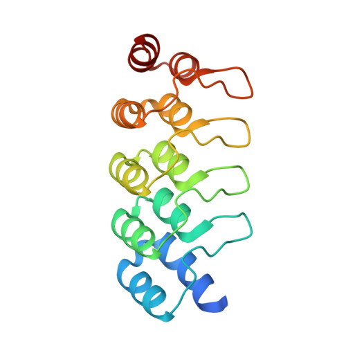
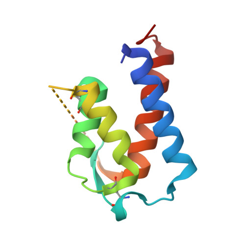
Conformational flexibility of an anti-IL-13 DARPin.

Teplyakov, A.Malia, T.J.Obmolova, G.Jacobs, S.A.O'Neil, K.T.Gilliland, G.L.

(2017) Protein Eng Des Sel 30: 31-37

  • DOI: https://doi.org/10.1093/protein/gzw059
  • Primary Citation Related Structures: 
    5KNG, 5KNH

  • PubMed Abstract: 

    Designed ankyrin repeat proteins (DARPin ® ) are artificial non-immunoglobulin binding proteins with potential applications as therapeutic molecules. DARPin 6G9 binds interleukin-13 with high affinity and blocks the signaling pathway and as such is promising for the treatment of asthma and other atopic diseases. The crystal structures of DARPin 6G9 in the unbound form and in complex with IL-13 were determined at high resolution. The DARPin competes for the same epitope as the IL-13 receptor chain 13Rα1 but does not interfere with the binding of the other receptor chain, IL-4Rα. Analysis of multiple copies of the DARPin molecule in the crystal indicates the conformational instability in the N-terminal cap that was predicted from molecular dynamics simulations. Comparison of the DARPin structures in the free state and in complex with IL-13 reveals a concerted movement of the ankyrin repeats upon binding resulted in the opening of the binding site. The induced-fit mode of binding employed by DARPin 6G9 is very unusual for DARPins since they were designed as particularly stable and rigid molecules. This finding shows that DARPins can operate by various binding mechanisms and suggests that some flexibility in the scaffold may be an advantage.


  • Organizational Affiliation
    • Janssen Research & Development, LLC, 1400 McKean Road, Spring House, PA 19477, USA ateplyak@its.jnj.com.

Macromolecule Content 

  • Total Structure Weight: 30.61 kDa 
  • Atom Count: 2,122 
  • Modeled Residue Count: 254 
  • Deposited Residue Count: 281 
  • Unique protein chains: 2

Macromolecules

Find similar proteins by:|  3D Structure
Entity ID: 1
MoleculeChains  Sequence LengthOrganismDetailsImage
DARPIN 6G9A [auth D]169synthetic constructMutation(s): 0 
Find similar proteins by:|  3D Structure
Entity ID: 2
MoleculeChains  Sequence LengthOrganismDetailsImage
IL13B [auth I]112Macaca fascicularisMutation(s): 0 
Gene Names: IL13
UniProt
Find proteins for Q0PW92 (Macaca fascicularis)
Explore Q0PW92 
Go to UniProtKB:  Q0PW92
Entity Groups
Sequence Clusters30% Identity50% Identity70% Identity90% Identity95% Identity100% Identity
UniProt GroupQ0PW92
Sequence Annotations
Expand
Reference Sequence

Small Molecules

Ligands 1 Unique
IDChains Name / Formula / InChI Key2D Diagram3D Interactions
ACT

Query on ACT


Download:Ideal Coordinates CCD File
C [auth D]ACETATE ION
C2 H3 O2
QTBSBXVTEAMEQO-UHFFFAOYSA-M

Experimental Data & Validation

Experimental Data

  • Method: X-RAY DIFFRACTION
  • Resolution: 1.60 Å
  • R-Value Free:  0.196 (Depositor), 0.202 (DCC) 
  • R-Value Work:  0.168 (Depositor) 
  • R-Value Observed: 0.169 (Depositor) 
Space Group: C 2 2 21
Unit Cell:
Length ( Å )Angle ( ˚ )
a = 52.41α = 90
b = 78.49β = 90
c = 119.44γ = 90
Software Package:
Software NamePurpose
XDSdata reduction
XDSdata scaling
PHASERphasing
REFMACrefinement

Structure Validation

View Full Validation Report


Entry History 

Deposition Data

Revision History  (Full details and data files)

  • Version 1.0: 2016-12-14
    Type: Initial release
  • Version 1.1: 2017-01-11
    Changes: Database references
  • Version 1.2: 2023-09-27
    Changes: Data collection, Database references, Refinement description
  • Version 1.3: 2024-11-13
    Changes: Structure summary