5KD4 | pdb_00005kd4

Crystal Structure of Murine MHC-I H-2Dd in complex with Murine Beta2-Microglobulin and a Variant of Peptide (PVI10) of HIV gp120 MN Isolate (IGPGRAFYVI)


Experimental Data Snapshot

  • Method: X-RAY DIFFRACTION
  • Resolution: 3.05 Å
  • R-Value Free: 
    0.224 (Depositor), 0.219 (DCC) 
  • R-Value Work: 
    0.184 (Depositor), 0.182 (DCC) 
  • R-Value Observed: 
    0.186 (Depositor) 

Starting Model: experimental
View more details

wwPDB Validation 3D Report Full Report

Validation slider image for 5KD4

This is version 1.4 of the entry. See complete history

Literature

Effects of Cross-Presentation, Antigen Processing, and Peptide Binding in HIV Evasion of T Cell Immunity.

Frey, B.F.Jiang, J.Sui, Y.Boyd, L.F.Yu, B.Tatsuno, G.Billeskov, R.Solaymani-Mohammadi, S.Berman, P.W.Margulies, D.H.Berzofsky, J.A.

(2018) J Immunol 200: 1853-1864

  • DOI: https://doi.org/10.4049/jimmunol.1701523
  • Primary Citation Related Structures: 
    5KD4, 5KD7, 5T7G

  • PubMed Abstract: 

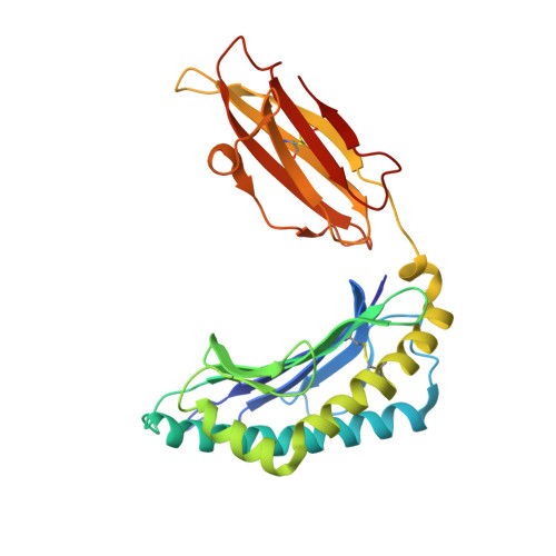
    Unlike cytosolic processing and presentation of viral Ags by virus-infected cells, Ags first expressed in infected nonprofessional APCs, such as CD4 + T cells in the case of HIV, are taken up by dendritic cells and cross-presented. This generally requires entry through the endocytic pathway, where endosomal proteases have first access for processing. Thus, understanding virus escape during cross-presentation requires an understanding of resistance to endosomal proteases, such as cathepsin S (CatS). We have modified HIV-1 MN gp120 by mutating a key CatS cleavage site (Thr 322 Thr 323 ) in the V3 loop of the immunodominant epitope IGPGRAFY TT to IGPGRAFY VV to prevent digestion. We found this mutation to facilitate cross-presentation and provide evidence from MHC binding and X-ray crystallographic structural studies that this results from preservation of the epitope rather than an increased epitope affinity for the MHC class I molecule. In contrast, when the protein is expressed by a vaccinia virus in the cytosol, the wild-type protein is immunogenic without this mutation. These proof-of-concept results show that a virus like HIV, infecting predominantly nonprofessional presenting cells, can escape T cell recognition by incorporating a CatS cleavage site that leads to destruction of an immunodominant epitope when the Ag undergoes endosomal cross-presentation.


  • Organizational Affiliation
    • Vaccine Branch, Center for Cancer Research, National Cancer Institute, National Institutes of Health, Bethesda, MD 20892.

Macromolecule Content 

  • Total Structure Weight: 90.3 kDa 
  • Atom Count: 6,278 
  • Modeled Residue Count: 764 
  • Deposited Residue Count: 774 
  • Unique protein chains: 3

Macromolecules

Find similar proteins by:|  3D Structure
Entity ID: 1
MoleculeChains  Sequence LengthOrganismDetailsImage
H-2 class I histocompatibility antigen, D-D alpha chainA,
D [auth C]
277Mus musculusMutation(s): 0 
Gene Names: H2-D1
UniProt
Find proteins for P01900 (Mus musculus)
Explore P01900 
Go to UniProtKB:  P01900
Entity Groups
Sequence Clusters30% Identity50% Identity70% Identity90% Identity95% Identity100% Identity
UniProt GroupP01900
Sequence Annotations
Expand
Reference Sequence
Find similar proteins by:|  3D Structure
Entity ID: 2
MoleculeChains  Sequence LengthOrganismDetailsImage
Beta-2-microglobulinB,
E [auth D]
100Mus musculusMutation(s): 0 
Gene Names: B2m
UniProt & NIH Common Fund Data Resources
Find proteins for P01887 (Mus musculus)
Explore P01887 
Go to UniProtKB:  P01887
IMPC:  MGI:88127
Entity Groups
Sequence Clusters30% Identity50% Identity70% Identity90% Identity95% Identity100% Identity
UniProt GroupP01887
Sequence Annotations
Expand
Reference Sequence
Find similar proteins by:  Sequence   |   3D Structure  
Entity ID: 3
MoleculeChains  Sequence LengthOrganismDetailsImage
Peptide of HIV gp120 MN isolate, PVI10 (IGPGRAFYVI)C [auth P],
F [auth Q]
10Human immunodeficiency virus 1Mutation(s): 0 
Gene Names: ENV
UniProt
Find proteins for P05877 (Human immunodeficiency virus type 1 group M subtype B (isolate MN))
Explore P05877 
Go to UniProtKB:  P05877
Entity Groups
Sequence Clusters30% Identity50% Identity70% Identity90% Identity95% Identity100% Identity
UniProt GroupP05877
Sequence Annotations
Expand
Reference Sequence

Experimental Data & Validation

Experimental Data

  • Method: X-RAY DIFFRACTION
  • Resolution: 3.05 Å
  • R-Value Free:  0.224 (Depositor), 0.219 (DCC) 
  • R-Value Work:  0.184 (Depositor), 0.182 (DCC) 
  • R-Value Observed: 0.186 (Depositor) 
Space Group: P 1 21 1
Unit Cell:
Length ( Å )Angle ( ˚ )
a = 47.13α = 90
b = 90.3β = 113.19
c = 120.07γ = 90
Software Package:
Software NamePurpose
PHENIXrefinement
XDSdata reduction
XDSdata scaling
PHASERphasing

Structure Validation

View Full Validation Report


Entry History 

Deposition Data

Revision History  (Full details and data files)

  • Version 1.0: 2017-10-11
    Type: Initial release
  • Version 1.1: 2018-02-07
    Changes: Database references
  • Version 1.2: 2018-02-28
    Changes: Database references
  • Version 1.3: 2023-09-27
    Changes: Data collection, Database references, Refinement description
  • Version 1.4: 2024-11-20
    Changes: Structure summary