5K8D

Crystal structure of rFVIIIFc


Experimental Data Snapshot

  • Method: X-RAY DIFFRACTION
  • Resolution: 4.19 Å
  • R-Value Free: 0.362 
  • R-Value Work: 0.298 
  • R-Value Observed: 0.301 

wwPDB Validation   3D Report Full Report


This is version 2.1 of the entry. See complete history


Literature

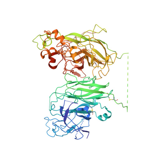
The structural basis for the functional comparability of factor VIII and the long-acting variant recombinant factor VIII Fc fusion protein.

Leksa, N.C.Chiu, P.L.Bou-Assaf, G.M.Quan, C.Liu, Z.Goodman, A.B.Chambers, M.G.Tsutakawa, S.E.Hammel, M.Peters, R.T.Walz, T.Kulman, J.D.

(2017) J Thromb Haemost 15: 1167-1179

  • DOI: https://doi.org/10.1111/jth.13700
  • Primary Citation of Related Structures:  
    5K8D

  • PubMed Abstract: 

    Essentials Recombinant factor VIII (rFVIII) Fc fusion protein has a 1.5-fold longer half-life than rFVIII. Five orthogonal methods were used to characterize the structure of rFVIIIFc compared to rFVIII. The C-terminal Fc fusion does not perturb the structure of FVIII in rFVIIIFc. The FVIII and Fc components of rFVIIIFc are flexibly tethered and functionally independent. Background Fusion of the human IgG 1 Fc domain to the C-terminal C2 domain of B-domain-deleted (BDD) factor VIII (FVIII) results in the recombinant FVIII Fc (rFVIIIFc) fusion protein, which has a 1.5-fold longer half-life in humans. Objective To assess the structural properties of rFVIIIFc by comparing its constituent FVIII and Fc elements with their respective isolated components, and evaluating their structural independence within rFVIIIFc. Methods rFVIIIFc and its isolated FVIII and Fc components were compared by the use of hydrogen-deuterium exchange mass spectrometry (HDX-MS). The structure of rFVIIIFc was also evaluated by the use of X-ray crystallography, small-angle X-ray scattering (SAXS), and electron microscopy (EM). The degree of steric interference by the appended Fc domain was assessed by EM and surface plasmon resonance (SPR). Results HDX-MS analysis of rFVIIIFc revealed that fusion caused no structural perturbations in FVIII or Fc. The rFVIIIFc crystal structure showed that the FVIII component is indistinguishable from published BDD FVIII structures. The Fc domain was not observed, indicating high mobility. SAXS analysis was consistent with an ensemble of rigid-body models in which the Fc domain exists in a largely extended orientation relative to FVIII. Binding of Fab fragments of anti-C2 domain antibodies to BDD FVIII was visualized by EM, and the affinities of the corresponding intact antibodies for BDD FVIII and rFVIIIFc were comparable by SPR analysis. Conclusions The FVIII and Fc components of rFVIIIFc are structurally indistinguishable from their isolated constituents, and show a high degree of structural independence, consistent with the functional comparability of rFVIIIFc and unmodified FVIII.


  • Organizational Affiliation

    Biogen, Cambridge, MA, USA.


Macromolecules
Find similar proteins by:  (by identity cutoff)  |  3D Structure
Entity ID: 1
MoleculeChains Sequence LengthOrganismDetailsImage
Coagulation factor VIII740Homo sapiensMutation(s): 0 
Gene Names: F8F8C
UniProt & NIH Common Fund Data Resources
Find proteins for P00451 (Homo sapiens)
Explore P00451 
Go to UniProtKB:  P00451
PHAROS:  P00451
GTEx:  ENSG00000185010 
Entity Groups  
Sequence Clusters30% Identity50% Identity70% Identity90% Identity95% Identity100% Identity
UniProt GroupP00451
Sequence Annotations
Expand
  • Reference Sequence
Find similar proteins by:  (by identity cutoff)  |  3D Structure
Entity ID: 2
MoleculeChains Sequence LengthOrganismDetailsImage
Coagulation factor VIII,Ig gamma-1 chain C region865Homo sapiensMutation(s): 0 
Gene Names: F8F8CIGHG1
UniProt & NIH Common Fund Data Resources
Find proteins for P01857 (Homo sapiens)
Explore P01857 
Go to UniProtKB:  P01857
PHAROS:  P01857
GTEx:  ENSG00000211896 
Find proteins for P00451 (Homo sapiens)
Explore P00451 
Go to UniProtKB:  P00451
PHAROS:  P00451
GTEx:  ENSG00000185010 
Entity Groups  
Sequence Clusters30% Identity50% Identity70% Identity90% Identity95% Identity100% Identity
UniProt GroupsP01857P00451
Sequence Annotations
Expand
  • Reference Sequence
Oligosaccharides

Help

Entity ID: 3
MoleculeChains Length2D Diagram Glycosylation3D Interactions
alpha-D-mannopyranose-(1-6)-beta-D-mannopyranose-(1-4)-2-acetamido-2-deoxy-beta-D-glucopyranose-(1-4)-2-acetamido-2-deoxy-beta-D-glucopyranose
C
4N-Glycosylation
Glycosylation Resources
GlyTouCan:  G22573RC
GlyCosmos:  G22573RC
GlyGen:  G22573RC
Entity ID: 4
MoleculeChains Length2D Diagram Glycosylation3D Interactions
2-acetamido-2-deoxy-beta-D-glucopyranose-(1-4)-2-acetamido-2-deoxy-beta-D-glucopyranose
D
2N-Glycosylation
Glycosylation Resources
GlyTouCan:  G42666HT
GlyCosmos:  G42666HT
GlyGen:  G42666HT
Entity ID: 5
MoleculeChains Length2D Diagram Glycosylation3D Interactions
alpha-D-mannopyranose-(1-2)-beta-D-mannopyranose-(1-3)-[alpha-D-mannopyranose-(1-6)-alpha-D-mannopyranose-(1-6)]beta-D-mannopyranose-(1-4)-2-acetamido-2-deoxy-beta-D-glucopyranose-(1-4)-2-acetamido-2-deoxy-beta-D-glucopyranose
E
7N-Glycosylation
Glycosylation Resources
GlyTouCan:  G71177VF
GlyCosmos:  G71177VF
GlyGen:  G71177VF
Experimental Data & Validation

Experimental Data

  • Method: X-RAY DIFFRACTION
  • Resolution: 4.19 Å
  • R-Value Free: 0.362 
  • R-Value Work: 0.298 
  • R-Value Observed: 0.301 
  • Space Group: P 41 21 2
Unit Cell:
Length ( Å )Angle ( ˚ )
a = 136.295α = 90
b = 136.295β = 90
c = 365.098γ = 90
Software Package:
Software NamePurpose
REFMACrefinement
SCALEPACKdata scaling
PHASERphasing
PDB_EXTRACTdata extraction
HKL-2000data reduction

Structure Validation

View Full Validation Report


Entry History 

Deposition Data

Revision History  (Full details and data files)

  • Version 1.0: 2017-06-14
    Type: Initial release
  • Version 1.1: 2017-06-28
    Changes: Database references
  • Version 2.0: 2020-07-29
    Type: Remediation
    Reason: Carbohydrate remediation
    Changes: Atomic model, Data collection, Derived calculations, Structure summary
  • Version 2.1: 2023-09-27
    Changes: Data collection, Database references, Derived calculations, Refinement description, Structure summary