5K5E | pdb_00005k5e

Discovery and Structure-Activity Relationships of a Highly Selective Butyrylcholinesterase Inhibitor by Structure-Based Virtual Screening


Experimental Data Snapshot

  • Method: X-RAY DIFFRACTION
  • Resolution: 2.80 Å
  • R-Value Free: 
    0.202 (Depositor), 0.202 (DCC) 
  • R-Value Work: 
    0.173 (Depositor), 0.175 (DCC) 
  • R-Value Observed: 
    0.175 (Depositor) 

Starting Model: experimental
View more details

wwPDB Validation 3D Report Full Report

Validation slider image for 5K5E

Ligand Structure Quality Assessment 


This is version 2.4 of the entry. See complete history

Literature

Discovery and Structure-Activity Relationships of a Highly Selective Butyrylcholinesterase Inhibitor by Structure-Based Virtual Screening.

Dighe, S.N.Deora, G.S.De la Mora, E.Nachon, F.Chan, S.Parat, M.O.Brazzolotto, X.Ross, B.P.

(2016) J Med Chem 59: 7683-7689

  • DOI: https://doi.org/10.1021/acs.jmedchem.6b00356
  • Primary Citation Related Structures: 
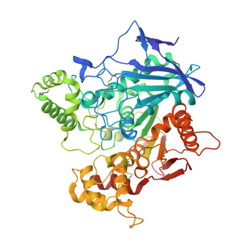
    5K5E

  • PubMed Abstract: 

    Structure-based virtual screening of two libraries containing 567 981 molecules was used to discover novel, selective BuChE inhibitors, which are potentially superior symptomatic treatments in late-stage Alzheimer's disease. Compound 16 was identified as a highly selective submicromolar inhibitor of BuChE (huBuChE IC50 = 0.443 μM) with high permeability in the PAMPA-BBB model. The X-ray crystal structure of huBuChE in complex with 16 revealed the atomic-level interactions and offers opportunities for further development of the series.


  • Organizational Affiliation
    • School of Pharmacy, The University of Queensland , Brisbane, Queensland 4072, Australia.

Macromolecule Content 

  • Total Structure Weight: 124.14 kDa 
  • Atom Count: 8,862 
  • Modeled Residue Count: 1,053 
  • Deposited Residue Count: 1,058 
  • Unique protein chains: 1

Macromolecules

Find similar proteins by:|  3D Structure
Entity ID: 1
MoleculeChains  Sequence LengthOrganismDetailsImage
Cholinesterase
A, B
529Homo sapiensMutation(s): 0 
Gene Names: BCHECHE1
EC: 3.1.1.8
UniProt & NIH Common Fund Data Resources
Find proteins for P06276 (Homo sapiens)
Explore P06276 
Go to UniProtKB:  P06276
PHAROS:  P06276
GTEx:  ENSG00000114200 
Entity Groups
Sequence Clusters30% Identity50% Identity70% Identity90% Identity95% Identity100% Identity
UniProt GroupP06276
Glycosylation
Glycosylation Sites: 6Go to GlyGen: P06276-1
Sequence Annotations
Expand
Reference Sequence

Oligosaccharides

Help  
Entity ID: 2
MoleculeChains Length2D Diagram GlycosylationD Interactions
2-acetamido-2-deoxy-beta-D-glucopyranose-(1-4)-2-acetamido-2-deoxy-beta-D-glucopyranose
C, D, E
2N-Glycosylation
Glycosylation Resources
GlyTouCan: G42666HT
GlyCosmos: G42666HT
GlyGen: G42666HT

Small Molecules

Ligands 4 Unique
IDChains Name / Formula / InChI Key2D Diagram3D Interactions
6QS

Query on 6QS


Download:Ideal Coordinates CCD File
J [auth A],
R [auth B]
[(2~{R})-oxolan-2-yl]methyl 4-(9-ethylcarbazol-3-yl)-2-methyl-5-oxidanylidene-4,6,7,8-tetrahydro-1~{H}-quinoline-3-carboxylate
C30 H32 N2 O4
SHBFRAVLHSPWTB-NGOKVRLYSA-N
NAG

Query on NAG


Download:Ideal Coordinates CCD File
F [auth A]
G [auth A]
H [auth A]
I [auth A]
L [auth B]
F [auth A],
G [auth A],
H [auth A],
I [auth A],
L [auth B],
M [auth B],
N [auth B],
O [auth B],
P [auth B],
Q [auth B]
2-acetamido-2-deoxy-beta-D-glucopyranose
C8 H15 N O6
OVRNDRQMDRJTHS-FMDGEEDCSA-N
GOL

Query on GOL


Download:Ideal Coordinates CCD File
U [auth B],
V [auth B]
GLYCEROL
C3 H8 O3
PEDCQBHIVMGVHV-UHFFFAOYSA-N
EDO

Query on EDO


Download:Ideal Coordinates CCD File
K [auth A],
S [auth B],
T [auth B]
1,2-ETHANEDIOL
C2 H6 O2
LYCAIKOWRPUZTN-UHFFFAOYSA-N

Experimental Data & Validation

Experimental Data

  • Method: X-RAY DIFFRACTION
  • Resolution: 2.80 Å
  • R-Value Free:  0.202 (Depositor), 0.202 (DCC) 
  • R-Value Work:  0.173 (Depositor), 0.175 (DCC) 
  • R-Value Observed: 0.175 (Depositor) 
Space Group: P 21 21 21
Unit Cell:
Length ( Å )Angle ( ˚ )
a = 75.81α = 90
b = 80.07β = 90
c = 230.63γ = 90
Software Package:
Software NamePurpose
PHENIXrefinement
XDSdata reduction
XSCALEdata scaling
PHASERphasing

Structure Validation

View Full Validation Report


Ligand Structure Quality Assessment 


Entry History 

Deposition Data

Revision History  (Full details and data files)

  • Version 1.0: 2016-07-27
    Type: Initial release
  • Version 1.1: 2016-09-07
    Changes: Database references
  • Version 1.2: 2018-05-09
    Changes: Data collection, Source and taxonomy
  • Version 1.3: 2019-02-20
    Changes: Advisory, Data collection, Derived calculations
  • Version 1.4: 2019-10-02
    Changes: Data collection, Derived calculations
  • Version 2.0: 2020-07-29
    Type: Remediation
    Reason: Carbohydrate remediation
    Changes: Advisory, Atomic model, Data collection, Derived calculations, Structure summary
  • Version 2.1: 2023-02-15
    Changes: Database references, Structure summary
  • Version 2.2: 2023-03-01
    Changes: Advisory
  • Version 2.3: 2024-02-07
    Changes: Data collection, Refinement description
  • Version 2.4: 2024-11-20
    Changes: Structure summary