5K3J | pdb_00005k3j

Crystals structure of Acyl-CoA oxidase-2 in Caenorhabditis elegans bound with FAD, ascaroside-CoA, and ATP


Experimental Data Snapshot

  • Method: X-RAY DIFFRACTION
  • Resolution: 2.68 Å
  • R-Value Free: 
    0.245 (Depositor), 0.262 (DCC) 
  • R-Value Work: 
    0.213 (Depositor), 0.229 (DCC) 
  • R-Value Observed: 
    0.215 (Depositor) 

Starting Model: experimental
View more details

wwPDB Validation 3D Report Full Report

Validation slider image for 5K3J

Ligand Structure Quality Assessment 


This is version 1.6 of the entry. See complete history

Literature

Structural characterization of acyl-CoA oxidases reveals a direct link between pheromone biosynthesis and metabolic state in Caenorhabditis elegans.

Zhang, X.Li, K.Jones, R.A.Bruner, S.D.Butcher, R.A.

(2016) Proc Natl Acad Sci U S A 113: 10055-10060

  • DOI: https://doi.org/10.1073/pnas.1608262113
  • Primary Citation Related Structures: 
    5K3G, 5K3H, 5K3I, 5K3J

  • PubMed Abstract: 

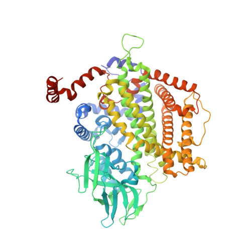
    Caenorhabditis elegans secretes ascarosides as pheromones to communicate with other worms and to coordinate the development and behavior of the population. Peroxisomal β-oxidation cycles shorten the side chains of ascaroside precursors to produce the short-chain ascaroside pheromones. Acyl-CoA oxidases, which catalyze the first step in these β-oxidation cycles, have different side chain-length specificities and enable C. elegans to regulate the production of specific ascaroside pheromones. Here, we determine the crystal structure of the acyl-CoA oxidase 1 (ACOX-1) homodimer and the ACOX-2 homodimer bound to its substrate. Our results provide a molecular basis for the substrate specificities of the acyl-CoA oxidases and reveal why some of these enzymes have a very broad substrate range, whereas others are quite specific. Our results also enable predictions to be made for the roles of uncharacterized acyl-CoA oxidases in C. elegans and in other nematode species. Remarkably, we show that most of the C. elegans acyl-CoA oxidases that participate in ascaroside biosynthesis contain a conserved ATP-binding pocket that lies at the dimer interface, and we identify key residues in this binding pocket. ATP binding induces a structural change that is associated with tighter binding of the FAD cofactor. Mutations that disrupt ATP binding reduce FAD binding and reduce enzyme activity. Thus, ATP may serve as a regulator of acyl-CoA oxidase activity, thereby directly linking ascaroside biosynthesis to ATP concentration and metabolic state.


  • Organizational Affiliation
    • Department of Chemistry, University of Florida, Gainesville, FL 32611.

Macromolecule Content 

  • Total Structure Weight: 158.1 kDa 
  • Atom Count: 10,979 
  • Modeled Residue Count: 1,321 
  • Deposited Residue Count: 1,348 
  • Unique protein chains: 1

Macromolecules

Find similar proteins by:|  3D Structure
Entity ID: 1
MoleculeChains  Sequence LengthOrganismDetailsImage
Acyl-coenzyme A oxidase
A, B
674Caenorhabditis elegansMutation(s): 0 
Gene Names: acox-2CELE_F08A8.2F08A8.2
EC: 1.3.3
UniProt
Find proteins for O62137 (Caenorhabditis elegans)
Explore O62137 
Go to UniProtKB:  O62137
Entity Groups
Sequence Clusters30% Identity50% Identity70% Identity90% Identity95% Identity100% Identity
UniProt GroupO62137
Sequence Annotations
Expand
Reference Sequence

Small Molecules

Ligands 4 Unique
IDChains Name / Formula / InChI Key2D Diagram3D Interactions
6QA

Query on 6QA


Download:Ideal Coordinates CCD File
E [auth A],
J [auth B]
~{S}-[2-[3-[[(2~{R})-4-[[[(2~{R},3~{S},4~{R},5~{R})-5-(6-aminopurin-9-yl)-4-oxidanyl-3-phosphonooxy-oxolan-2-yl]methoxy-oxidanyl-phosphoryl]oxy-oxidanyl-phosphoryl]oxy-3,3-dimethyl-2-oxidanyl-butanoyl]amino]propanoylamino]ethyl] 5-[(2~{R},3~{R},5~{R},6~{S})-6-methyl-3,5-bis(oxidanyl)oxan-2-yl]oxypentanethioate
C32 H54 N7 O21 P3 S
OJJVYBQJWBBERR-XKGQWQQESA-N
FAD

Query on FAD


Download:Ideal Coordinates CCD File
C [auth A],
G [auth B]
FLAVIN-ADENINE DINUCLEOTIDE
C27 H33 N9 O15 P2
VWWQXMAJTJZDQX-UYBVJOGSSA-N
ATP

Query on ATP


Download:Ideal Coordinates CCD File
D [auth A],
H [auth B]
ADENOSINE-5'-TRIPHOSPHATE
C10 H16 N5 O13 P3
ZKHQWZAMYRWXGA-KQYNXXCUSA-N
MG

Query on MG


Download:Ideal Coordinates CCD File
F [auth B],
I [auth B]
MAGNESIUM ION
Mg
JLVVSXFLKOJNIY-UHFFFAOYSA-N

Experimental Data & Validation

Experimental Data

  • Method: X-RAY DIFFRACTION
  • Resolution: 2.68 Å
  • R-Value Free:  0.245 (Depositor), 0.262 (DCC) 
  • R-Value Work:  0.213 (Depositor), 0.229 (DCC) 
  • R-Value Observed: 0.215 (Depositor) 
Space Group: P 1 21 1
Unit Cell:
Length ( Å )Angle ( ˚ )
a = 76.04α = 90
b = 85.571β = 91.43
c = 106.899γ = 90
Software Package:
Software NamePurpose
PHENIXrefinement
XDSdata reduction
Aimlessdata scaling
PHASERphasing
BUCCANEERmodel building

Structure Validation

View Full Validation Report


Ligand Structure Quality Assessment 


Entry History 

& Funding Information

Deposition Data


Funding OrganizationLocationGrant Number
Research Corporation for Science AdvancementUnited States22844
Ellison Medical FoundationUnited StatesAG-NS-0963-12
National Science Foundation (NSF, United States)United States1555050
Alfred P. Sloan FoundationUnited StatesBR2014-071

Revision History  (Full details and data files)

  • Version 1.0: 2016-08-24
    Type: Initial release
  • Version 1.1: 2016-09-07
    Changes: Database references
  • Version 1.2: 2016-09-14
    Changes: Database references
  • Version 1.3: 2017-09-27
    Changes: Author supporting evidence, Database references, Derived calculations
  • Version 1.4: 2019-11-27
    Changes: Author supporting evidence
  • Version 1.5: 2024-03-06
    Changes: Data collection, Database references
  • Version 1.6: 2024-04-03
    Changes: Refinement description