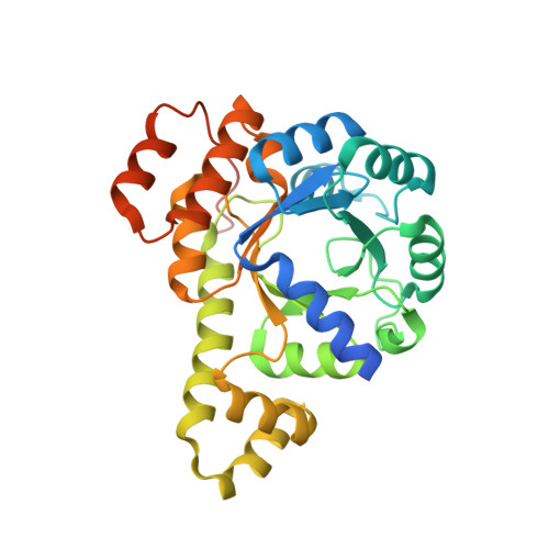
5K2Z

PDX1.3-adduct (Arabidopsis)


Experimental Data Snapshot

  • Method: X-RAY DIFFRACTION
  • Resolution: 1.80 Å
  • R-Value Free: 0.204 
  • R-Value Work: 0.180 
  • R-Value Observed: 0.181 

Starting Model: experimental
View more details

wwPDB Validation   3D Report Full Report


This is version 1.3 of the entry. See complete history


Literature

Structural definition of the lysine swing in Arabidopsis thaliana PDX1: Intermediate channeling facilitating vitamin B6 biosynthesis.

Robinson, G.C.Kaufmann, M.Roux, C.Fitzpatrick, T.B.

(2016) Proc Natl Acad Sci U S A 113: E5821-E5829

  • DOI: https://doi.org/10.1073/pnas.1608125113
  • Primary Citation of Related Structures:  
    5K2Z, 5K3V

  • PubMed Abstract: 

    Vitamin B 6 is indispensible for all organisms, notably as the coenzyme form pyridoxal 5'-phosphate. Plants make the compound de novo using a relatively simple pathway comprising pyridoxine synthase (PDX1) and pyridoxine glutaminase (PDX2). PDX1 is remarkable given its multifaceted synthetic ability to carry out isomerization, imine formation, ammonia addition, aldol-type condensation, cyclization, and aromatization, all in the absence of coenzymes or recruitment of specialized domains. Two active sites (P1 and P2) facilitate the plethora of reactions, but it is not known how the two are coordinated and, moreover, if intermediates are tunneled between active sites. Here we present X-ray structures of PDX1.3 from Arabidopsis thaliana, the overall architecture of which is a dodecamer of (β/α) 8 barrels, similar to the majority of its homologs. An apoenzyme structure revealed that features around the P1 active site in PDX1.3 have adopted inward conformations consistent with a catalytically primed state and delineated a substrate accessible cavity above this active site, not noted in other reported structures. Comparison with the structure of PDX1.3 with an intermediate along the catalytic trajectory demonstrated that a lysine residue swings from the distinct P2 site to the P1 site at this stage of catalysis and is held in place by a molecular catch and pin, positioning it for transfer of serviced substrate back to P2. The study shows that a simple lysine swinging arm coordinates use of chemically disparate sites, dispensing with the need for additional factors, and provides an elegant example of solving complex chemistry to generate an essential metabolite.


  • Organizational Affiliation

    Department of Botany and Plant Biology, University of Geneva, 1211 Geneva, Switzerland.


Macromolecules
Find similar proteins by:  (by identity cutoff)  |  3D Structure
Entity ID: 1
MoleculeChains Sequence LengthOrganismDetailsImage
Pyridoxal 5'-phosphate synthase subunit PDX1.3
A, B, C, D
315Arabidopsis thalianaMutation(s): 0 
Gene Names: PDX13GIP2PDX1L3RSR4At5g01410T10O8.120
EC: 4.3.3.6
UniProt
Find proteins for Q8L940 (Arabidopsis thaliana)
Explore Q8L940 
Go to UniProtKB:  Q8L940
Entity Groups  
Sequence Clusters30% Identity50% Identity70% Identity90% Identity95% Identity100% Identity
UniProt GroupQ8L940
Sequence Annotations
Expand
  • Reference Sequence
Small Molecules
Ligands 4 Unique
IDChains Name / Formula / InChI Key2D Diagram3D Interactions
6R3
Query on 6R3

Download Ideal Coordinates CCD File 
H [auth A],
M [auth B],
Q [auth C],
V [auth D]
2-azanylpenta-1,4-dien-3-one
C5 H7 N O
AHTXUWSCLWCTPN-UHFFFAOYSA-N
SO4
Query on SO4

Download Ideal Coordinates CCD File 
F [auth A]
G [auth A]
K [auth B]
L [auth B]
O [auth C]
F [auth A],
G [auth A],
K [auth B],
L [auth B],
O [auth C],
P [auth C],
T [auth D],
U [auth D]
SULFATE ION
O4 S
QAOWNCQODCNURD-UHFFFAOYSA-L
EDO
Query on EDO

Download Ideal Coordinates CCD File 
I [auth A],
J [auth A],
N [auth B],
R [auth C]
1,2-ETHANEDIOL
C2 H6 O2
LYCAIKOWRPUZTN-UHFFFAOYSA-N
CL
Query on CL

Download Ideal Coordinates CCD File 
E [auth A],
S [auth D]
CHLORIDE ION
Cl
VEXZGXHMUGYJMC-UHFFFAOYSA-M
Experimental Data & Validation

Experimental Data

  • Method: X-RAY DIFFRACTION
  • Resolution: 1.80 Å
  • R-Value Free: 0.204 
  • R-Value Work: 0.180 
  • R-Value Observed: 0.181 
  • Space Group: H 3
Unit Cell:
Length ( Å )Angle ( ˚ )
a = 178.46α = 90
b = 178.46β = 90
c = 115.81γ = 120
Software Package:
Software NamePurpose
MOSFLMdata reduction
SCALAdata scaling
PHASERphasing
REFMACrefinement
PDB_EXTRACTdata extraction
PHASERphasing

Structure Validation

View Full Validation Report


Entry History & Funding Information

Deposition Data


Funding OrganizationLocationGrant Number
Swiss National Science FoundationSwitzerland31003A-141117/1

Revision History  (Full details and data files)

  • Version 1.0: 2016-10-05
    Type: Initial release
  • Version 1.1: 2016-10-19
    Changes: Database references
  • Version 1.2: 2024-01-10
    Changes: Data collection, Database references, Refinement description
  • Version 1.3: 2024-11-20
    Changes: Structure summary