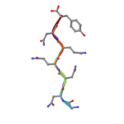
5K2H

Structure of GNNQQNY from yeast prion Sup35 in space group P212121 determined by MicroED

  • Classification: PROTEIN FIBRIL
  • Organism(s): Saccharomyces cerevisiae
  • Mutation(s): No 

  • Deposited: 2016-05-18 Released: 2016-09-14 
  • Deposition Author(s): Rodriguez, J.A., Sawaya, M.R., Cascio, D., Eisenberg, D.S.
  • Funding Organization(s): National Science Foundation (NSF, United States), National Institutes of Health/National Institute on Aging (NIH/NIA), Alzheimer's Disease Reasearch Center, Howard Hughes Medical Institute (HHMI), Department of Energy (DOE, United States), Giannini Foundation

Experimental Data Snapshot

  • Method: ELECTRON CRYSTALLOGRAPHY
  • Resolution: 1.05 Å
  • R-Value Free: 0.186 
  • R-Value Work: 0.177 
  • R-Value Observed: 0.178 

wwPDB Validation   3D Report Full Report

Currently 5K2H does not have a validation slider image.


This is version 1.9 of the entry. See complete history


Literature

Ab initio structure determination from prion nanocrystals at atomic resolution by MicroED.

Sawaya, M.R.Rodriguez, J.Cascio, D.Collazo, M.J.Shi, D.Reyes, F.E.Hattne, J.Gonen, T.Eisenberg, D.S.

(2016) Proc Natl Acad Sci U S A 113: 11232-11236

  • DOI: https://doi.org/10.1073/pnas.1606287113
  • Primary Citation of Related Structures:  
    5K2E, 5K2F, 5K2G, 5K2H

  • PubMed Abstract: 

    Electrons, because of their strong interaction with matter, produce high-resolution diffraction patterns from tiny 3D crystals only a few hundred nanometers thick in a frozen-hydrated state. This discovery offers the prospect of facile structure determination of complex biological macromolecules, which cannot be coaxed to form crystals large enough for conventional crystallography or cannot easily be produced in sufficient quantities. Two potential obstacles stand in the way. The first is a phenomenon known as dynamical scattering, in which multiple scattering events scramble the recorded electron diffraction intensities so that they are no longer informative of the crystallized molecule. The second obstacle is the lack of a proven means of de novo phase determination, as is required if the molecule crystallized is insufficiently similar to one that has been previously determined. We show with four structures of the amyloid core of the Sup35 prion protein that, if the diffraction resolution is high enough, sufficiently accurate phases can be obtained by direct methods with the cryo-EM method microelectron diffraction (MicroED), just as in X-ray diffraction. The success of these four experiments dispels the concern that dynamical scattering is an obstacle to ab initio phasing by MicroED and suggests that structures of novel macromolecules can also be determined by direct methods.


  • Organizational Affiliation

    Howard Hughes Medical Institute, University of California, Los Angeles, CA 90024-1570; University of California, Los Angeles-Department of Energy Institute, University of California, Los Angeles, CA 90024-1570; Department of Biological Chemistry, University of California, Los Angeles, CA 90024-1570; Department of Chemistry and Biochemistry, University of California, Los Angeles, CA 90024-1570; Molecular Biology Institute, University of California, Los Angeles, CA 90024-1570.


Macromolecules

Find similar proteins by:  Sequence   |   3D Structure  

Entity ID: 1
MoleculeChains Sequence LengthOrganismDetailsImage
Eukaryotic peptide chain release factor GTP-binding subunit7Saccharomyces cerevisiaeMutation(s): 0 
EC: 3.6.5
UniProt
Find proteins for P05453 (Saccharomyces cerevisiae (strain ATCC 204508 / S288c))
Explore P05453 
Go to UniProtKB:  P05453
Entity Groups  
UniProt GroupP05453
Sequence Annotations
Expand
  • Reference Sequence
Experimental Data & Validation

Experimental Data

  • Method: ELECTRON CRYSTALLOGRAPHY
  • Resolution: 1.05 Å
  • R-Value Free: 0.186 
  • R-Value Work: 0.177 
  • R-Value Observed: 0.178 
  • Space Group: P 21 21 21
Unit Cell:
Length ( Å )Angle ( ˚ )
a = 23.16α = 90
b = 4.93β = 90
c = 40.51γ = 90
Software Package:
Software NamePurpose
XSCALEdata scaling
REFMACrefinement
PDB_EXTRACTdata extraction
EM Software:
TaskSoftware PackageVersion
MODEL REFINEMENTrefmac55.8.0135
RECONSTRUCTIONSHELXD2013/2

Structure Validation

View Full Validation Report

Currently 5K2H does not have a validation slider image.


Entry History & Funding Information

Deposition Data


Funding OrganizationLocationGrant Number
National Science Foundation (NSF, United States)United StatesMCB-0445429
National Institutes of Health/National Institute on Aging (NIH/NIA)United States1R01-AG029430
Alzheimer's Disease Reasearch CenterUnited States--
Howard Hughes Medical Institute (HHMI)United States--
Department of Energy (DOE, United States)United StatesDE-FC02-02ER63421
Giannini FoundationUnited States--

Revision History  (Full details and data files)

  • Version 1.0: 2016-09-14
    Type: Initial release
  • Version 1.1: 2016-09-21
    Changes: Database references
  • Version 1.2: 2016-10-05
    Changes: Database references
  • Version 1.3: 2016-10-19
    Changes: Database references
  • Version 1.4: 2016-11-30
    Changes: Refinement description
  • Version 1.5: 2017-09-13
    Changes: Author supporting evidence, Data collection
  • Version 1.6: 2018-04-25
    Changes: Data collection
  • Version 1.7: 2019-11-20
    Changes: Author supporting evidence
  • Version 1.8: 2021-06-30
    Changes: Data collection
  • Version 1.9: 2024-03-06
    Changes: Data collection, Database references