5JYG | pdb_00005jyg

Cryo-EM structure of the MamK filament at 6.5 A


Experimental Data Snapshot

  • Method: ELECTRON MICROSCOPY
  • Resolution: 6.50 Å
  • Aggregation State: FILAMENT 
  • Reconstruction Method: HELICAL 

wwPDB Validation 3D Report Full Report

Validation slider image for 5JYG

This is version 1.2 of the entry. See complete history

Literature

Structure of the magnetosome-associated actin-like MamK filament at subnanometer resolution.

Bergeron, J.R.Hutto, R.Ozyamak, E.Hom, N.Hansen, J.Draper, O.Byrne, M.E.Keyhani, S.Komeili, A.Kollman, J.M.

(2017) Protein Sci 26: 93-102

  • DOI: https://doi.org/10.1002/pro.2979
  • Primary Citation Related Structures: 
    5JYG

  • PubMed Abstract: 

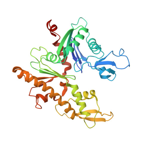
    Magnetotactic bacteria possess cellular compartments called magnetosomes that sense magnetic fields. Alignment of magnetosomes in the bacterial cell is necessary for their function, and this is achieved through anchoring of magnetosomes to filaments composed of the protein MamK. MamK is an actin homolog that polymerizes upon ATP binding. Here, we report the structure of the MamK filament at ∼6.5 Å, obtained by cryo-Electron Microscopy. This structure confirms our previously reported double-stranded, nonstaggered architecture, and reveals the molecular basis for filament formation. While MamK is closest in sequence to the bacterial actin MreB, the longitudinal contacts along each MamK strand most closely resemble those of eukaryotic actin. In contrast, the cross-strand interface, with a surprisingly limited set of contacts, is novel among actin homologs and gives rise to the nonstaggered architecture.


  • Organizational Affiliation
    • Department of Biochemistry, University of Washington, Seattle, Washington.

Macromolecule Content 

  • Total Structure Weight: 533.51 kDa 
  • Atom Count: 35,462 
  • Modeled Residue Count: 4,592 
  • Deposited Residue Count: 4,858 
  • Unique protein chains: 1

Macromolecules

Find similar proteins by:|  3D Structure
Entity ID: 1
MoleculeChains  Sequence LengthOrganismDetailsImage
Actin-like ATPase
A, B, C, D, E
A, B, C, D, E, F, G, H, I, J, K, L, M, N
347Paramagnetospirillum magneticum AMB-1Mutation(s): 0 
Gene Names: amb0965
EC: 3.6.1
UniProt
Find proteins for Q2W8Q6 (Paramagnetospirillum magneticum (strain ATCC 700264 / AMB-1))
Explore Q2W8Q6 
Go to UniProtKB:  Q2W8Q6
Entity Groups
Sequence Clusters30% Identity50% Identity70% Identity90% Identity95% Identity100% Identity
UniProt GroupQ2W8Q6
Sequence Annotations
Expand
Reference Sequence

Small Molecules

Ligands 2 Unique
IDChains Name / Formula / InChI Key2D Diagram3D Interactions
ADP

Query on ADP


Download:Ideal Coordinates CCD File
BA [auth G]
DA [auth H]
FA [auth I]
HA [auth J]
JA [auth K]
BA [auth G],
DA [auth H],
FA [auth I],
HA [auth J],
JA [auth K],
LA [auth L],
NA [auth M],
P [auth A],
PA [auth N],
R [auth B],
T [auth C],
V [auth D],
X [auth E],
Z [auth F]
ADENOSINE-5'-DIPHOSPHATE
C10 H15 N5 O10 P2
XTWYTFMLZFPYCI-KQYNXXCUSA-N
MG

Query on MG


Download:Ideal Coordinates CCD File
AA [auth G]
CA [auth H]
EA [auth I]
GA [auth J]
IA [auth K]
AA [auth G],
CA [auth H],
EA [auth I],
GA [auth J],
IA [auth K],
KA [auth L],
MA [auth M],
O [auth A],
OA [auth N],
Q [auth B],
S [auth C],
U [auth D],
W [auth E],
Y [auth F]
MAGNESIUM ION
Mg
JLVVSXFLKOJNIY-UHFFFAOYSA-N

Experimental Data & Validation

Experimental Data

  • Method: ELECTRON MICROSCOPY
  • Resolution: 6.50 Å
  • Aggregation State: FILAMENT 
  • Reconstruction Method: HELICAL 
EM Software:
TaskSoftware PackageVersion
MODEL REFINEMENTRosetta3.5

Structure Validation

View Full Validation Report


Entry History 

Deposition Data

Revision History  (Full details and data files)

  • Version 1.0: 2016-07-27
    Type: Initial release
  • Version 1.1: 2017-01-11
    Changes: Database references
  • Version 1.2: 2024-03-06
    Changes: Data collection, Database references