5JVX | pdb_00005jvx

X-ray structure of the adduct formed in the reaction between thaumatin and a gold carbene compound


Experimental Data Snapshot

  • Method: X-RAY DIFFRACTION
  • Resolution: 1.70 Å
  • R-Value Free: 
    0.187 (Depositor), 0.188 (DCC) 
  • R-Value Work: 
    0.170 (Depositor), 0.171 (DCC) 
  • R-Value Observed: 
    0.171 (Depositor) 

Starting Model: experimental
View more details

wwPDB Validation 3D Report Full Report

Validation slider image for 5JVX

Ligand Structure Quality Assessment 


This is version 1.3 of the entry. See complete history

Literature

First Crystal Structure for a Gold Carbene-Protein Adduct.

Ferraro, G.Gabbiani, C.Merlino, A.

(2016) Bioconjug Chem 27: 1584-1587

  • DOI: https://doi.org/10.1021/acs.bioconjchem.6b00298
  • Primary Citation Related Structures: 
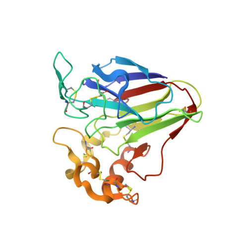
    5JVX

  • PubMed Abstract: 

    The X-ray structure of the adduct formed in the reaction between the gold N-heterocyclic carbene compound Au(NHC)Cl (with NHC = 1-butyl-3-methyl-imidazole-2-ylidene) and the model protein thaumatin is reported here. The structure reveals binding of Au(NHC)(+) fragments to distinct protein sites. Notably, binding of the gold compound occurs at lysine side chains and at the N-terminal tail; the metal binds the protein after releasing Cl(-) ligand, but retaining NHC fragment.


  • Organizational Affiliation
    • Department of Chemical Sciences, University of Naples Federico II, Complesso Universitario di Monte Sant'Angelo , Via Cintia, I-80126 Napoli, Italy.

Macromolecule Content 

  • Total Structure Weight: 24.79 kDa 
  • Atom Count: 2,020 
  • Modeled Residue Count: 207 
  • Deposited Residue Count: 207 
  • Unique protein chains: 1

Macromolecules

Find similar proteins by:|  3D Structure
Entity ID: 1
MoleculeChains  Sequence LengthOrganismDetailsImage
Thaumatin-1207Thaumatococcus danielliiMutation(s): 0 
UniProt
Find proteins for P02883 (Thaumatococcus daniellii)
Explore P02883 
Go to UniProtKB:  P02883
Entity Groups
Sequence Clusters30% Identity50% Identity70% Identity90% Identity95% Identity100% Identity
UniProt GroupP02883
Sequence Annotations
Expand
Reference Sequence

Small Molecules

Ligands 3 Unique
IDChains Name / Formula / InChI Key2D Diagram3D Interactions
6O0

Query on 6O0


Download:Ideal Coordinates CCD File
D [auth A]
E [auth A]
F [auth A]
G [auth A]
H [auth A]
D [auth A],
E [auth A],
F [auth A],
G [auth A],
H [auth A],
I [auth A]
(1-butyl-3-methyl-1,3-dihydro-2H-imidazol-2-ylidene)(chloro)gold
C8 H14 Au Cl N2
VPJMTYLCIKLAAU-UHFFFAOYSA-M
TLA

Query on TLA


Download:Ideal Coordinates CCD File
B [auth A],
C [auth A]
L(+)-TARTARIC ACID
C4 H6 O6
FEWJPZIEWOKRBE-JCYAYHJZSA-N
CL

Query on CL


Download:Ideal Coordinates CCD File
J [auth A]CHLORIDE ION
Cl
VEXZGXHMUGYJMC-UHFFFAOYSA-M

Experimental Data & Validation

Experimental Data

  • Method: X-RAY DIFFRACTION
  • Resolution: 1.70 Å
  • R-Value Free:  0.187 (Depositor), 0.188 (DCC) 
  • R-Value Work:  0.170 (Depositor), 0.171 (DCC) 
  • R-Value Observed: 0.171 (Depositor) 
Space Group: P 41 21 2
Unit Cell:
Length ( Å )Angle ( ˚ )
a = 57.912α = 90
b = 57.912β = 90
c = 150.136γ = 90
Software Package:
Software NamePurpose
REFMACrefinement
HKL-2000data reduction
HKL-2000data scaling
PHASERphasing

Structure Validation

View Full Validation Report


Ligand Structure Quality Assessment 


Entry History 

Deposition Data

Revision History  (Full details and data files)

  • Version 1.0: 2016-07-13
    Type: Initial release
  • Version 1.1: 2016-07-27
    Changes: Database references
  • Version 1.2: 2024-01-10
    Changes: Advisory, Data collection, Database references, Refinement description
  • Version 1.3: 2024-10-16
    Changes: Structure summary