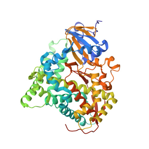
5JTD

Crystal structure of the Ru(bpy)2PhenA functionalized P450 BM3 L407C heme domain mutant in complex with DMSO.


Experimental Data Snapshot

  • Method: X-RAY DIFFRACTION
  • Resolution: 1.50 Å
  • R-Value Free: 0.198 
  • R-Value Work: 0.173 
  • R-Value Observed: 0.174 

Starting Model: experimental
View more details

wwPDB Validation   3D Report Full Report


Ligand Structure Quality Assessment 


This is version 1.2 of the entry. See complete history


Literature

Insights into an efficient light-driven hybrid P450 BM3 enzyme from crystallographic, spectroscopic and biochemical studies.

Spradlin, J.Lee, D.Mahadevan, S.Mahomed, M.Tang, L.Lam, Q.Colbert, A.Shafaat, O.S.Goodin, D.Kloos, M.Kato, M.Cheruzel, L.E.

(2016) Biochim Biophys Acta 1864: 1732-1738

  • DOI: https://doi.org/10.1016/j.bbapap.2016.09.005
  • Primary Citation of Related Structures:  
    5JQ2, 5JTD

  • PubMed Abstract: 

    In order to perform selective CH functionalization upon visible light irradiation, Ru(II)-diimine functionalized P450 heme enzymes have been developed. The sL407C-1 enzyme containing the Ru(bpy) 2 PhenA (bpy=2,2'-bipyridine and PhenA=5-acetamido-1,10-phenanthroline) photosensitizer (1) covalently attached to the non-native single cysteine L407C of the P450BM3 heme domain mutant, displays high photocatalytic activity in the selective CH bond hydroxylation of several substrates. A combination of X-ray crystallography, site-directed mutagenesis, transient absorption measurements and enzymatic assays was used to gain insights into its photocatalytic activity and electron transfer pathway. The crystal structure of the sL407C-1 enzyme was solved in the open and closed conformations revealing a through-space electron transfer pathway involving highly conserved, F393 and Q403, residues. Several mutations of these residues (F393A, F393W or Q403W) were introduced to probe their roles in the overall reaction. Transient absorption measurements confirm rapid electron transfer as heme reduction is observed in all four hybrid enzymes. Compared to the parent sL407C-1, photocatalytic activity was negligible in the dF393A-1 enzyme while 60% increase in activity with total turnover numbers of 420 and 90% product conversion was observed with the dQ403W-1 mutant. In the sL407C-1 enzyme, the photosensitizer is ideally located to rapidly deliver electrons, using the naturally occurring electron transfer pathway, to the heme center in order to activate molecular dioxygen and sustain photocatalytic activity. The results shed light on the design of efficient light-driven biocatalysts and the approach can be generalized to other members of the P450 superfamily.


  • Organizational Affiliation

    San José State University, Department of Chemistry, One Washington Square, San José, CA, United States.


Macromolecules
Find similar proteins by:  (by identity cutoff)  |  3D Structure
Entity ID: 1
MoleculeChains Sequence LengthOrganismDetailsImage
Bifunctional cytochrome P450/NADPH--P450 reductase
A, B
469Priestia megaterium NBRC 15308 = ATCC 14581Mutation(s): 1 
Gene Names: cyp102A1cyp102BG04_163
EC: 1.14.14.1 (PDB Primary Data), 1.6.2.4 (PDB Primary Data)
UniProt
Find proteins for P14779 (Priestia megaterium (strain ATCC 14581 / DSM 32 / CCUG 1817 / JCM 2506 / NBRC 15308 / NCIMB 9376 / NCTC 10342 / NRRL B-14308 / VKM B-512 / Ford 19))
Explore P14779 
Go to UniProtKB:  P14779
Entity Groups  
Sequence Clusters30% Identity50% Identity70% Identity90% Identity95% Identity100% Identity
UniProt GroupP14779
Sequence Annotations
Expand
  • Reference Sequence
Small Molecules
Ligands 3 Unique
IDChains Name / Formula / InChI Key2D Diagram3D Interactions
RU8
Query on RU8

Download Ideal Coordinates CCD File 
E [auth A],
I [auth B]
bis(2,2'-bipyridine-kappa~2~N~1~,N~1'~)[2-iodo-N-(1,10-phenanthrolin-5-yl-kappa~2~N~1~,N~10~)acetamide]ruthenium(2+)
C34 H26 I N7 O Ru
IMVNBPJSKRZLRT-UHFFFAOYSA-N
HEM
Query on HEM

Download Ideal Coordinates CCD File 
C [auth A],
G [auth B]
PROTOPORPHYRIN IX CONTAINING FE
C34 H32 Fe N4 O4
KABFMIBPWCXCRK-RGGAHWMASA-L
DMS
Query on DMS

Download Ideal Coordinates CCD File 
D [auth A],
F [auth A],
H [auth B],
J [auth B]
DIMETHYL SULFOXIDE
C2 H6 O S
IAZDPXIOMUYVGZ-UHFFFAOYSA-N
Experimental Data & Validation

Experimental Data

  • Method: X-RAY DIFFRACTION
  • Resolution: 1.50 Å
  • R-Value Free: 0.198 
  • R-Value Work: 0.173 
  • R-Value Observed: 0.174 
  • Space Group: P 1 21 1
Unit Cell:
Length ( Å )Angle ( ˚ )
a = 58.69α = 90
b = 145.39β = 97.08
c = 62.9γ = 90
Software Package:
Software NamePurpose
REFMACrefinement
XSCALEdata scaling
PHASERphasing
PDB_EXTRACTdata extraction
XDSdata reduction

Structure Validation

View Full Validation Report


Ligand Structure Quality Assessment 


Entry History 

Deposition Data

  • Released Date: 2016-10-12 
  • Deposition Author(s): Kloos, M.

Revision History  (Full details and data files)

  • Version 1.0: 2016-10-12
    Type: Initial release
  • Version 1.1: 2023-09-27
    Changes: Data collection, Database references, Derived calculations, Refinement description
  • Version 1.2: 2024-10-16
    Changes: Structure summary