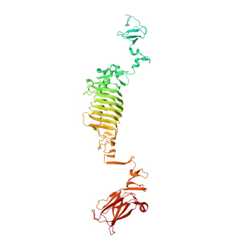
5JSE

Crystal structure of phiAB6 tailspike in complex with three-repeated oligosaccharides of Acinetobacter baumannii surface polysaccharide


Experimental Data Snapshot

  • Method: X-RAY DIFFRACTION
  • Resolution: 1.89 Å
  • R-Value Free: 0.165 
  • R-Value Work: 0.131 
  • R-Value Observed: 0.132 

wwPDB Validation   3D Report Full Report


This is version 3.1 of the entry. See complete history


Literature

Structural basis for fragmenting the exopolysaccharide of Acinetobacter baumannii by bacteriophage Phi AB6 tailspike protein

Lee, I.M.Tu, I.F.Yang, F.L.Ko, T.P.Liao, J.H.Lin, N.T.Wu, C.Y.Ren, C.T.Wang, A.H.Chang, C.M.Huang, K.F.Wu, S.H.

(2017) Sci Rep 7: 42711-42711

  • DOI: https://doi.org/10.1038/srep42711
  • Primary Citation of Related Structures:  
    5JS4, 5JSD, 5JSE

  • PubMed Abstract: 

    With an increase in antibiotic-resistant strains, the nosocomial pathogen Acinetobacter baumannii has become a serious threat to global health. Glycoconjugate vaccines containing fragments of bacterial exopolysaccharide (EPS) are an emerging therapeutic to combat bacterial infection. Herein, we characterize the bacteriophage ΦAB6 tailspike protein (TSP), which specifically hydrolyzed the EPS of A. baumannii strain 54149 (Ab-54149). Ab-54149 EPS exhibited the same chemical structure as two antibiotic-resistant A. baumannii strains. The ΦAB6 TSP-digested products comprised oligosaccharides of two repeat units, typically with stoichiometric pseudaminic acid (Pse). The 1.48-1.89-Å resolution crystal structures of an N-terminally-truncated ΦAB6 TSP and its complexes with the semi-hydrolyzed products revealed a trimeric β-helix architecture that bears intersubunit carbohydrate-binding grooves, with some features unusual to the TSP family. The structures suggest that Pse in the substrate is an important recognition site for ΦAB6 TSP. A region in the carbohydrate-binding groove is identified as the determinant of product specificity. The structures also elucidated a retaining mechanism, for which the catalytic residues were verified by site-directed mutagenesis. Our findings provide a structural basis for engineering the enzyme to produce desired oligosaccharides, which is useful for the development of glycoconjugate vaccines against A. baumannii infection.


  • Organizational Affiliation

    Institute of Biochemical Sciences, National Taiwan University, Taipei 106, Taiwan.


Macromolecules
Find similar proteins by:  (by identity cutoff)  |  3D Structure
Entity ID: 1
MoleculeChains Sequence LengthOrganismDetailsImage
phiAB6 tailspike
A, B, C
719Bacteriophage sp.Mutation(s): 0 
UniProt
Find proteins for A0A159BDB5 (Acinetobacter phage phiAB6)
Explore A0A159BDB5 
Go to UniProtKB:  A0A159BDB5
Entity Groups  
Sequence Clusters30% Identity50% Identity70% Identity90% Identity95% Identity100% Identity
UniProt GroupA0A159BDB5
Sequence Annotations
Expand
  • Reference Sequence
Oligosaccharides

Help

Entity ID: 2
MoleculeChains Length2D Diagram Glycosylation3D Interactions
beta-D-galactopyranose-(1-3)-2-amino-2-deoxy-beta-D-galactopyranose-(1-3)-[5,7-bisacetamido-3,5,7,9-tetradeoxy-L-glycero-alpha-L-manno-non-2-ulopyranosonic acid-(2-6)-beta-D-glucopyranose-(1-6)]beta-D-galactopyranose-(1-3)-2-amino-2-deoxy-beta-D-galactopyranose-(1-3)-[beta-D-glucopyranose-(1-6)]beta-D-galactopyranose
D, E, F
8N/A
Glycosylation Resources
GlyTouCan:  G07591XG
GlyCosmos:  G07591XG
Small Molecules
Ligands 2 Unique
IDChains Name / Formula / InChI Key2D Diagram3D Interactions
MLA
Query on MLA

Download Ideal Coordinates CCD File 
G [auth A]
H [auth A]
K [auth B]
L [auth B]
O [auth C]
G [auth A],
H [auth A],
K [auth B],
L [auth B],
O [auth C],
P [auth C]
MALONIC ACID
C3 H4 O4
OFOBLEOULBTSOW-UHFFFAOYSA-N
ACY
Query on ACY

Download Ideal Coordinates CCD File 
I [auth A]
J [auth A]
M [auth B]
N [auth B]
Q [auth C]
I [auth A],
J [auth A],
M [auth B],
N [auth B],
Q [auth C],
R [auth C]
ACETIC ACID
C2 H4 O2
QTBSBXVTEAMEQO-UHFFFAOYSA-N
Experimental Data & Validation

Experimental Data

  • Method: X-RAY DIFFRACTION
  • Resolution: 1.89 Å
  • R-Value Free: 0.165 
  • R-Value Work: 0.131 
  • R-Value Observed: 0.132 
  • Space Group: C 1 2 1
Unit Cell:
Length ( Å )Angle ( ˚ )
a = 135.519α = 90
b = 78.139β = 100.47
c = 248.405γ = 90
Software Package:
Software NamePurpose
REFMACrefinement
HKL-2000data reduction
HKL-2000data scaling
PHENIXphasing

Structure Validation

View Full Validation Report


Entry History & Funding Information

Deposition Data


Funding OrganizationLocationGrant Number
Ministry of Science and Technology and Academia SinicaTaiwanNSC101-2923-B-001-005-MY3, MOST104-2325-B-001-003, NSC-101-2319-B-001-003, and MOST105-0210-01-12-01

Revision History  (Full details and data files)

  • Version 1.0: 2017-03-08
    Type: Initial release
  • Version 2.0: 2017-11-29
    Changes: Advisory, Atomic model, Data collection, Derived calculations, Non-polymer description, Refinement description, Structure summary
  • Version 3.0: 2020-07-29
    Type: Remediation
    Reason: Carbohydrate remediation
    Changes: Advisory, Atomic model, Data collection, Derived calculations, Structure summary
  • Version 3.1: 2024-03-20
    Changes: Data collection, Database references, Derived calculations, Structure summary