5JO8 | pdb_00005jo8

CEP104 TOG domain


Experimental Data Snapshot

  • Method: X-RAY DIFFRACTION
  • Resolution: 1.40 Å
  • R-Value Free: 
    0.238 (Depositor), 0.239 (DCC) 
  • R-Value Work: 
    0.219 (Depositor), 0.220 (DCC) 
  • R-Value Observed: 
    0.219 (Depositor) 

wwPDB Validation 3D Report Full Report

Validation slider image for 5JO8

This is version 1.3 of the entry. See complete history

Literature

Biophysical and Structural Characterization of the Centriolar Protein Cep104 Interaction Network.

Rezabkova, L.Kraatz, S.H.Akhmanova, A.Steinmetz, M.O.Kammerer, R.A.

(2016) J Biological Chem 291: 18496-18504

  • DOI: https://doi.org/10.1074/jbc.M116.739771
  • Primary Citation Related Structures: 
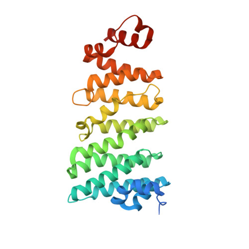
    5JO8

  • PubMed Abstract: 

    Dysfunction of cilia is associated with common genetic disorders termed ciliopathies. Knowledge on the interaction networks of ciliary proteins is therefore key for understanding the processes that are underlying these severe diseases and the mechanisms of ciliogenesis in general. Cep104 has recently been identified as a key player in the regulation of cilia formation. Using a combination of sequence analysis, biophysics, and x-ray crystallography, we obtained new insights into the domain architecture and interaction network of the Cep104 protein. We solved the crystal structure of the tumor overexpressed gene (TOG) domain, identified Cep104 as a novel tubulin-binding protein, and biophysically characterized the interaction of Cep104 with CP110, Cep97, end-binding (EB) protein, and tubulin. Our results represent a solid platform for the further investigation of the microtubule-EB-Cep104-tubulin-CP110-Cep97 network of proteins. Ultimately, such studies should be of importance for understanding the process of cilia formation and the mechanisms underlying different ciliopathies.


  • Organizational Affiliation
    • From the Laboratory of Biomolecular Research, Division of Biology and Chemistry, Paul Scherrer Institute, CH-5232 Villigen PSI, Switzerland and.

Macromolecule Content 

  • Total Structure Weight: 31.38 kDa 
  • Atom Count: 2,321 
  • Modeled Residue Count: 256 
  • Deposited Residue Count: 279 
  • Unique protein chains: 1

Macromolecules

Find similar proteins by:|  3D Structure
Entity ID: 1
MoleculeChains  Sequence LengthOrganismDetailsImage
CEP104279Gallus gallusMutation(s): 0 
Gene Names: CEP104

Experimental Data & Validation

Experimental Data

  • Method: X-RAY DIFFRACTION
  • Resolution: 1.40 Å
  • R-Value Free:  0.238 (Depositor), 0.239 (DCC) 
  • R-Value Work:  0.219 (Depositor), 0.220 (DCC) 
  • R-Value Observed: 0.219 (Depositor) 
Space Group: P 2 21 21
Unit Cell:
Length ( Å )Angle ( ˚ )
a = 48.36α = 90
b = 57.22β = 90
c = 102.96γ = 90
Software Package:
Software NamePurpose
PHENIXrefinement
XDSdata reduction
XSCALEdata scaling
AutoSolphasing
PHASERphasing

Structure Validation

View Full Validation Report


Entry History 

& Funding Information

Deposition Data


Funding OrganizationLocationGrant Number
Marie Curie IEFBelgium--
European Molecular Biology OrganizationGermany--

Revision History  (Full details and data files)

  • Version 1.0: 2016-07-20
    Type: Initial release
  • Version 1.1: 2016-09-07
    Changes: Database references
  • Version 1.2: 2017-09-06
    Changes: Author supporting evidence
  • Version 1.3: 2024-05-08
    Changes: Data collection, Database references