5JGE

Crystal structure of Atg19 coiled-coil complexed with Ape1 propeptide


Experimental Data Snapshot

  • Method: X-RAY DIFFRACTION
  • Resolution: 1.91 Å
  • R-Value Free: 0.246 
  • R-Value Work: 0.194 
  • R-Value Observed: 0.197 

wwPDB Validation   3D Report Full Report


This is version 1.3 of the entry. See complete history


Literature

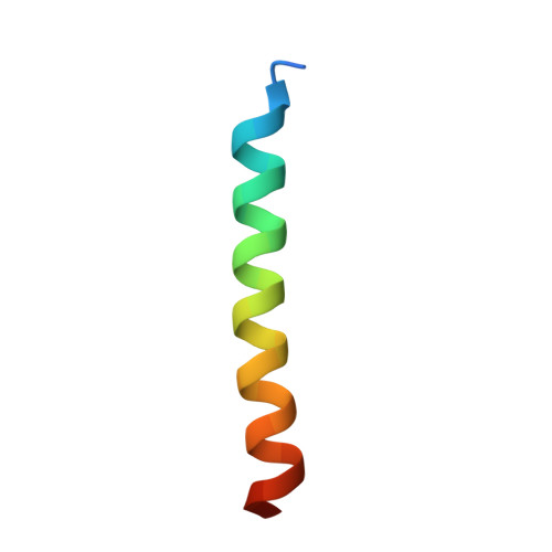
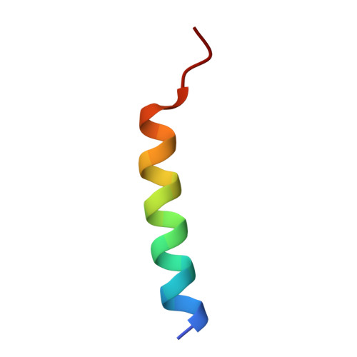
Structural Basis for Receptor-Mediated Selective Autophagy of Aminopeptidase I Aggregates

Yamasaki, A.Watanabe, Y.Adachi, W.Suzuki, K.Matoba, K.Kirisako, H.Kumeta, H.Nakatogawa, H.Ohsumi, Y.Inagaki, F.Noda, N.N.

(2016) Cell Rep 16: 19-27

  • DOI: https://doi.org/10.1016/j.celrep.2016.05.066
  • Primary Citation of Related Structures:  
    5JGE, 5JGF, 5JH9, 5JHC

  • PubMed Abstract: 

    Selective autophagy mediates the degradation of various cargoes, including protein aggregates and organelles, thereby contributing to cellular homeostasis. Cargo receptors ensure selectivity by tethering specific cargo to lipidated Atg8 at the isolation membrane. However, little is known about the structural requirements underlying receptor-mediated cargo recognition. Here, we report structural, biochemical, and cell biological analysis of the major selective cargo protein in budding yeast, aminopeptidase I (Ape1), and its complex with the receptor Atg19. The Ape1 propeptide has a trimeric coiled-coil structure, which tethers dodecameric Ape1 bodies together to form large aggregates. Atg19 disassembles the propeptide trimer and forms a 2:1 heterotrimer, which not only blankets the Ape1 aggregates but also regulates their size. These receptor activities may promote elongation of the isolation membrane along the aggregate surface, enabling sequestration of the cargo with high specificity.


  • Organizational Affiliation

    Institute of Microbial Chemistry (BIKAKEN), Tokyo 141-0021, Japan.


Macromolecules
Find similar proteins by:  (by identity cutoff)  |  3D Structure
Entity ID: 1
MoleculeChains Sequence LengthOrganismDetailsImage
Autophagy-related protein 19
A, B, D, E
32Saccharomyces cerevisiae S288CMutation(s): 0 
Gene Names: ATG19CVT19YOL082WO0980YOL01
UniProt
Find proteins for P35193 (Saccharomyces cerevisiae (strain ATCC 204508 / S288c))
Explore P35193 
Go to UniProtKB:  P35193
Entity Groups  
Sequence Clusters30% Identity50% Identity70% Identity90% Identity95% Identity100% Identity
UniProt GroupP35193
Sequence Annotations
Expand
  • Reference Sequence

Find similar proteins by:  Sequence   |   3D Structure  

Entity ID: 2
MoleculeChains Sequence LengthOrganismDetailsImage
Ape1 propeptide
C, F
23Saccharomyces cerevisiaeMutation(s): 0 
UniProt
Find proteins for P14904 (Saccharomyces cerevisiae (strain ATCC 204508 / S288c))
Explore P14904 
Go to UniProtKB:  P14904
Entity Groups  
Sequence Clusters30% Identity50% Identity70% Identity90% Identity95% Identity100% Identity
UniProt GroupP14904
Sequence Annotations
Expand
  • Reference Sequence
Experimental Data & Validation

Experimental Data

  • Method: X-RAY DIFFRACTION
  • Resolution: 1.91 Å
  • R-Value Free: 0.246 
  • R-Value Work: 0.194 
  • R-Value Observed: 0.197 
  • Space Group: P 1
Unit Cell:
Length ( Å )Angle ( ˚ )
a = 27.875α = 87.19
b = 32.921β = 76.52
c = 56.957γ = 67.1
Software Package:
Software NamePurpose
REFMACrefinement
HKL-2000data reduction
HKL-2000data scaling
PHENIXphasing

Structure Validation

View Full Validation Report


Entry History & Funding Information

Deposition Data


Funding OrganizationLocationGrant Number
Japan Science and Technology AgencyJapan--

Revision History  (Full details and data files)

  • Version 1.0: 2016-06-29
    Type: Initial release
  • Version 1.1: 2016-07-13
    Changes: Database references
  • Version 1.2: 2020-02-19
    Changes: Data collection, Derived calculations
  • Version 1.3: 2024-03-20
    Changes: Data collection, Database references, Refinement description