5JCZ | pdb_00005jcz

Rab11 bound to MyoVa-GTD


Experimental Data Snapshot

  • Method: X-RAY DIFFRACTION
  • Resolution: 2.06 Å
  • R-Value Free: 
    0.208 (Depositor), 0.212 (DCC) 
  • R-Value Work: 
    0.180 (Depositor), 0.185 (DCC) 
  • R-Value Observed: 
    0.181 (Depositor) 

Starting Model: experimental
View more details

wwPDB Validation 3D Report Full Report

Validation slider image for 5JCZ

Ligand Structure Quality Assessment 


This is version 2.1 of the entry. See complete history

Literature

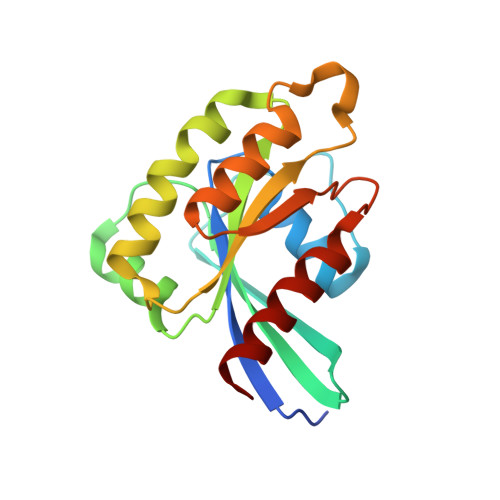
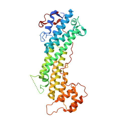
Coordinated recruitment of Spir actin nucleators and myosin V motors to Rab11 vesicle membranes.

Pylypenko, O.Welz, T.Tittel, J.Kollmar, M.Chardon, F.Malherbe, G.Weiss, S.Michel, C.I.Samol-Wolf, A.Grasskamp, A.T.Hume, A.Goud, B.Baron, B.England, P.Titus, M.A.Schwille, P.Weidemann, T.Houdusse, A.Kerkhoff, E.

(2016) Elife 5

  • DOI: https://doi.org/10.7554/eLife.17523
  • Primary Citation Related Structures: 
    5JCY, 5JCZ

  • PubMed Abstract: 

    There is growing evidence for a coupling of actin assembly and myosin motor activity in cells. However, mechanisms for recruitment of actin nucleators and motors on specific membrane compartments remain unclear. Here we report how Spir actin nucleators and myosin V motors coordinate their specific membrane recruitment. The myosin V globular tail domain (MyoV-GTD) interacts directly with an evolutionarily conserved Spir sequence motif. We determined crystal structures of MyoVa-GTD bound either to the Spir-2 motif or to Rab11 and show that a Spir-2:MyoVa:Rab11 complex can form. The ternary complex architecture explains how Rab11 vesicles support coordinated F-actin nucleation and myosin force generation for vesicle transport and tethering. New insights are also provided into how myosin activation can be coupled with the generation of actin tracks. Since MyoV binds several Rab GTPases, synchronized nucleator and motor targeting could provide a common mechanism to control force generation and motility in different cellular processes.


  • Organizational Affiliation
    • Institut Curie, PSL Research University, CNRS, UMR 144, F-75005, Paris, France.

Macromolecule Content 

  • Total Structure Weight: 199.09 kDa 
  • Atom Count: 13,842 
  • Modeled Residue Count: 1,581 
  • Deposited Residue Count: 1,728 
  • Unique protein chains: 2

Macromolecules

Find similar proteins by:|  3D Structure
Entity ID: 1
MoleculeChains  Sequence LengthOrganismDetailsImage
Ras-related protein Rab-11AA,
D,
F [auth I]
179Homo sapiensMutation(s): 0 
Gene Names: RAB11ARAB11
EC: 3.6.5.2
UniProt & NIH Common Fund Data Resources
Find proteins for P62491 (Homo sapiens)
Explore P62491 
Go to UniProtKB:  P62491
PHAROS:  P62491
GTEx:  ENSG00000103769 
Entity Groups
Sequence Clusters30% Identity50% Identity70% Identity90% Identity95% Identity100% Identity
UniProt GroupP62491
Sequence Annotations
Expand
Reference Sequence
Find similar proteins by:|  3D Structure
Entity ID: 2
MoleculeChains  Sequence LengthOrganismDetailsImage
Unconventional myosin-Va
B, C, E
397Homo sapiensMutation(s): 0 
Gene Names: MYO5AMYH12
UniProt & NIH Common Fund Data Resources
Find proteins for Q9Y4I1 (Homo sapiens)
Explore Q9Y4I1 
Go to UniProtKB:  Q9Y4I1
PHAROS:  Q9Y4I1
GTEx:  ENSG00000197535 
Entity Groups
Sequence Clusters30% Identity50% Identity70% Identity90% Identity95% Identity100% Identity
UniProt GroupQ9Y4I1
Sequence Annotations
Expand
Reference Sequence

Small Molecules

Ligands 6 Unique
IDChains Name / Formula / InChI Key2D Diagram3D Interactions
GDP

Query on GDP


Download:Ideal Coordinates CCD File
FA [auth I],
H [auth A],
Y [auth D]
GUANOSINE-5'-DIPHOSPHATE
C10 H15 N5 O11 P2
QGWNDRXFNXRZMB-UUOKFMHZSA-N
GOL

Query on GOL


Download:Ideal Coordinates CCD File
N [auth A],
Q [auth B]
GLYCEROL
C3 H8 O3
PEDCQBHIVMGVHV-UHFFFAOYSA-N
BEF

Query on BEF


Download:Ideal Coordinates CCD File
GA [auth I],
I [auth A],
Z [auth D]
BERYLLIUM TRIFLUORIDE ION
Be F3
OGIAHMCCNXDTIE-UHFFFAOYSA-K
EDO

Query on EDO


Download:Ideal Coordinates CCD File
AA [auth D]
BA [auth D]
CA [auth E]
DA [auth E]
J [auth A]
AA [auth D],
BA [auth D],
CA [auth E],
DA [auth E],
J [auth A],
K [auth A],
L [auth A],
O [auth B],
P [auth B],
R [auth C],
S [auth C],
T [auth C],
U [auth C]
1,2-ETHANEDIOL
C2 H6 O2
LYCAIKOWRPUZTN-UHFFFAOYSA-N
ACT

Query on ACT


Download:Ideal Coordinates CCD File
M [auth A],
W [auth C]
ACETATE ION
C2 H3 O2
QTBSBXVTEAMEQO-UHFFFAOYSA-M
MG

Query on MG


Download:Ideal Coordinates CCD File
EA [auth I],
G [auth A],
V [auth C],
X [auth D]
MAGNESIUM ION
Mg
JLVVSXFLKOJNIY-UHFFFAOYSA-N

Experimental Data & Validation

Experimental Data

  • Method: X-RAY DIFFRACTION
  • Resolution: 2.06 Å
  • R-Value Free:  0.208 (Depositor), 0.212 (DCC) 
  • R-Value Work:  0.180 (Depositor), 0.185 (DCC) 
  • R-Value Observed: 0.181 (Depositor) 
Space Group: C 1 2 1
Unit Cell:
Length ( Å )Angle ( ˚ )
a = 215.79α = 90
b = 128.42β = 98.27
c = 89.02γ = 90
Software Package:
Software NamePurpose
PHENIXrefinement
XDSdata reduction
XDSdata scaling
MOLREPphasing

Structure Validation

View Full Validation Report


Ligand Structure Quality Assessment 


Entry History 

Deposition Data

Revision History  (Full details and data files)

  • Version 1.0: 2016-09-28
    Type: Initial release
  • Version 2.0: 2018-10-24
    Changes: Atomic model, Data collection, Derived calculations
  • Version 2.1: 2024-01-10
    Changes: Data collection, Database references, Derived calculations, Refinement description