5JA8 | pdb_00005ja8

Crystal structure of the HigB2 toxin in complex with Nb2


Experimental Data Snapshot

  • Method: X-RAY DIFFRACTION
  • Resolution: 2.49 Å
  • R-Value Free: 
    0.248 (Depositor), 0.245 (DCC) 
  • R-Value Work: 
    0.201 (Depositor), 0.208 (DCC) 
  • R-Value Observed: 
    0.204 (Depositor) 

wwPDB Validation 3D Report Full Report

Validation slider image for 5JA8

This is version 1.3 of the entry. See complete history

Literature

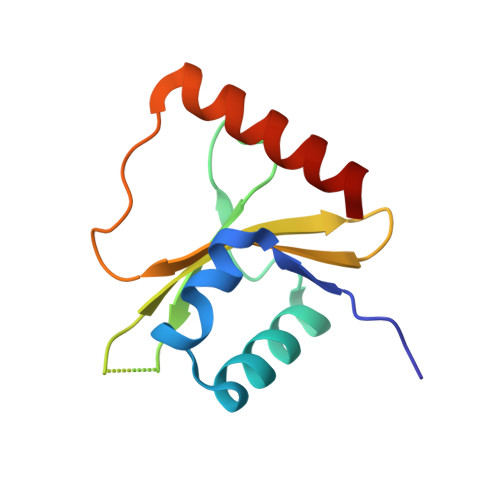
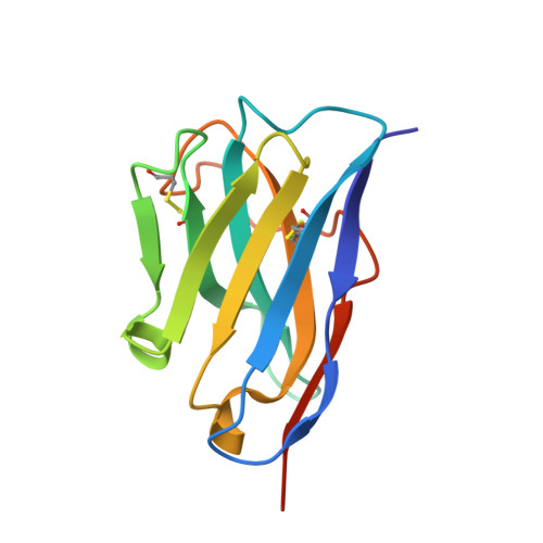
Ribosome-dependent Vibrio cholerae mRNAse HigB2 is regulated by a beta-strand sliding mechanism.

Hadzi, S.Garcia-Pino, A.Haesaerts, S.Jurenas, D.Gerdes, K.Lah, J.Loris, R.

(2017) Nucleic Acids Res 45: 4972-4983

  • DOI: https://doi.org/10.1093/nar/gkx138
  • Primary Citation Related Structures: 
    5J9I, 5JA8, 5JA9, 5JAA, 5MJE

  • PubMed Abstract: 

    Toxin-antitoxin (TA) modules are small operons involved in bacterial stress response and persistence. higBA operons form a family of TA modules with an inverted gene organization and a toxin belonging to the RelE/ParE superfamily. Here, we present the crystal structures of chromosomally encoded Vibrio cholerae antitoxin (VcHigA2), toxin (VcHigB2) and their complex, which show significant differences in structure and mechanisms of function compared to the higBA module from plasmid Rts1, the defining member of the family. The VcHigB2 is more closely related to Escherichia coli RelE both in terms of overall structure and the organization of its active site. VcHigB2 is neutralized by VcHigA2, a modular protein with an N-terminal intrinsically disordered toxin-neutralizing segment followed by a C-terminal helix-turn-helix dimerization and DNA binding domain. VcHigA2 binds VcHigB2 with picomolar affinity, which is mainly a consequence of entropically favorable de-solvation of a large hydrophobic binding interface and enthalpically favorable folding of the N-terminal domain into an α-helix followed by a β-strand. This interaction displaces helix α3 of VcHigB2 and at the same time induces a one-residue shift in the register of β-strand β3, thereby flipping the catalytically important Arg64 out of the active site.


  • Organizational Affiliation
    • Structural Biology Brussels, Department of Biotechnology, Vrije Universiteit Brussel, B-1050 Brussel, Belgium.

Macromolecule Content 

  • Total Structure Weight: 110.11 kDa 
  • Atom Count: 7,483 
  • Modeled Residue Count: 922 
  • Deposited Residue Count: 952 
  • Unique protein chains: 2

Macromolecules

Find similar proteins by:|  3D Structure
Entity ID: 1
MoleculeChains  Sequence LengthOrganismDetailsImage
Toxin HigB-2
A, C, E, G
111Vibrio cholerae O1 biovar El Tor str. N16961Mutation(s): 0 
Gene Names: higB-2VC_A0468
UniProt
Find proteins for Q9KMA6 (Vibrio cholerae serotype O1 (strain ATCC 39315 / El Tor Inaba N16961))
Explore Q9KMA6 
Go to UniProtKB:  Q9KMA6
Entity Groups
Sequence Clusters30% Identity50% Identity70% Identity90% Identity95% Identity100% Identity
UniProt GroupQ9KMA6
Sequence Annotations
Expand
Reference Sequence
Find similar proteins by:|  3D Structure
Entity ID: 2
MoleculeChains  Sequence LengthOrganismDetailsImage
Nanobody 2
B, D, F, H
127Lama glamaMutation(s): 0 

Small Molecules

Ligands 5 Unique
IDChains Name / Formula / InChI Key2D Diagram3D Interactions
EPE

Query on EPE


Download:Ideal Coordinates CCD File
P [auth D],
V [auth F]
4-(2-HYDROXYETHYL)-1-PIPERAZINE ETHANESULFONIC ACID
C8 H18 N2 O4 S
JKMHFZQWWAIEOD-UHFFFAOYSA-N
PO4

Query on PO4


Download:Ideal Coordinates CCD File
I [auth A]
J [auth A]
L [auth B]
N [auth C]
O [auth C]
I [auth A],
J [auth A],
L [auth B],
N [auth C],
O [auth C],
Q [auth D],
R [auth D],
T [auth E],
U [auth E],
W [auth G]
PHOSPHATE ION
O4 P
NBIIXXVUZAFLBC-UHFFFAOYSA-K
PDO

Query on PDO


Download:Ideal Coordinates CCD File
S [auth D],
X [auth G]
1,3-PROPANDIOL
C3 H8 O2
YPFDHNVEDLHUCE-UHFFFAOYSA-N
EDO

Query on EDO


Download:Ideal Coordinates CCD File
M [auth B]1,2-ETHANEDIOL
C2 H6 O2
LYCAIKOWRPUZTN-UHFFFAOYSA-N
ACT

Query on ACT


Download:Ideal Coordinates CCD File
K [auth A]ACETATE ION
C2 H3 O2
QTBSBXVTEAMEQO-UHFFFAOYSA-M

Experimental Data & Validation

Experimental Data

  • Method: X-RAY DIFFRACTION
  • Resolution: 2.49 Å
  • R-Value Free:  0.248 (Depositor), 0.245 (DCC) 
  • R-Value Work:  0.201 (Depositor), 0.208 (DCC) 
  • R-Value Observed: 0.204 (Depositor) 
Space Group: P 21 21 21
Unit Cell:
Length ( Å )Angle ( ˚ )
a = 76α = 90
b = 109.28β = 90
c = 117.48γ = 90
Software Package:
Software NamePurpose
XSCALEdata scaling
PHENIXrefinement
PDB_EXTRACTdata extraction
XDSdata reduction
PHASERphasing

Structure Validation

View Full Validation Report


Entry History 

Deposition Data

Revision History  (Full details and data files)

  • Version 1.0: 2017-04-05
    Type: Initial release
  • Version 1.1: 2017-05-17
    Changes: Database references
  • Version 1.2: 2019-02-20
    Changes: Advisory, Data collection, Derived calculations
  • Version 1.3: 2024-10-23
    Changes: Data collection, Database references, Refinement description, Structure summary