5J6X | pdb_00005j6x

Crystal structure of the apo-Zalpha of Zebrafish PKZ


Experimental Data Snapshot

  • Method: X-RAY DIFFRACTION
  • Resolution: 2.49 Å
  • R-Value Free: 
    0.269 (Depositor), 0.243 (DCC) 
  • R-Value Work: 
    0.182 (Depositor), 0.195 (DCC) 
  • R-Value Observed: 
    0.192 (Depositor) 

Starting Model: experimental
View more details

wwPDB Validation 3D Report Full Report

Validation slider image for 5J6X

This is version 1.2 of the entry. See complete history

Literature

Structural and functional studies of a large winged Z-DNA-binding domain of Danio rerio protein kinase PKZ

Subramani, V.K.Kim, D.Yun, K.Kim, K.K.

(2016) FEBS Lett 590: 2275-2285

  • DOI: https://doi.org/10.1002/1873-3468.12238
  • Primary Citation Related Structures: 
    5J6X

  • PubMed Abstract: 

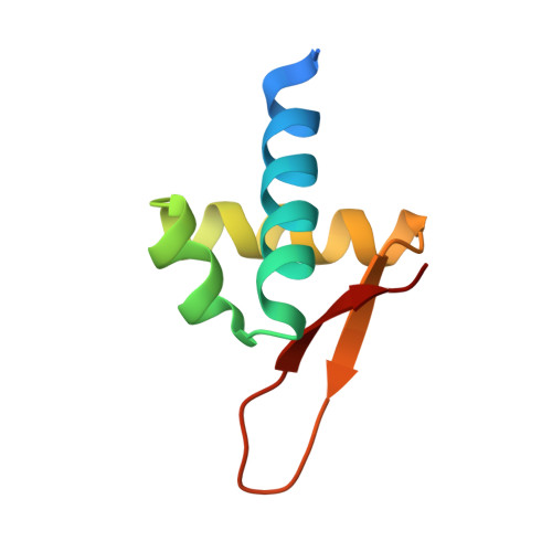
    The Z-DNA-binding domain of PKZ from zebrafish (Danio rerio; drZαPKZ ) contains the largest β-wing among known Z-DNA-binding domains. To elucidate the functional implication of the β-wing, we solved the crystal structure of apo-drZαPKZ . Structural comparison with its Z-DNA-bound form revealed a large conformational change within the β-wing during Z-DNA binding. Biochemical studies of protein mutants revealed that two basic residues in the β-wing are responsible for Z-DNA recognition as well as fast B-Z transition. Therefore, the extra basic residues in the β-wing of drZαPKZ are necessary for the fast B-Z transition activity.


  • Organizational Affiliation
    • Department of Molecular Cell Biology, Sungkyunkwan University School of Medicine, Suwon, Korea.

Macromolecule Content 

  • Total Structure Weight: 17.08 kDa 
  • Atom Count: 1,117 
  • Modeled Residue Count: 131 
  • Deposited Residue Count: 144 
  • Unique protein chains: 1

Macromolecules

Find similar proteins by:|  3D Structure
Entity ID: 1
MoleculeChains  Sequence LengthOrganismDetailsImage
Z-DNA binding protein kinase
A, B
72Danio rerioMutation(s): 0 
Gene Names: pkz
UniProt
Find proteins for Q5NE12 (Danio rerio)
Explore Q5NE12 
Go to UniProtKB:  Q5NE12
Entity Groups
Sequence Clusters30% Identity50% Identity70% Identity90% Identity95% Identity100% Identity
UniProt GroupQ5NE12
Sequence Annotations
Expand
Reference Sequence

Experimental Data & Validation

Experimental Data

  • Method: X-RAY DIFFRACTION
  • Resolution: 2.49 Å
  • R-Value Free:  0.269 (Depositor), 0.243 (DCC) 
  • R-Value Work:  0.182 (Depositor), 0.195 (DCC) 
  • R-Value Observed: 0.192 (Depositor) 
Space Group: P 41
Unit Cell:
Length ( Å )Angle ( ˚ )
a = 44.304α = 90
b = 44.304β = 90
c = 71.369γ = 90
Software Package:
Software NamePurpose
PHENIXrefinement
HKL-2000data reduction
HKL-2000data scaling
MOLREPphasing

Structure Validation

View Full Validation Report


Entry History 

& Funding Information

Deposition Data


Funding OrganizationLocationGrant Number
Samsung Science & Technology FoundationKorea, Republic OfSSTF-BA1301-01

Revision History  (Full details and data files)

  • Version 1.0: 2016-09-07
    Type: Initial release
  • Version 1.1: 2021-09-15
    Changes: Author supporting evidence, Database references, Derived calculations
  • Version 1.2: 2023-11-08
    Changes: Data collection, Refinement description