5J6F | pdb_00005j6f

Crystal structure of DAH7PS-CM complex from Geobacillus sp. with prephenate


Experimental Data Snapshot

  • Method: X-RAY DIFFRACTION
  • Resolution: 2.75 Å
  • R-Value Free: 
    0.235 (Depositor), 0.232 (DCC) 
  • R-Value Work: 
    0.182 (Depositor), 0.186 (DCC) 
  • R-Value Observed: 
    0.185 (Depositor) 

Starting Model: experimental
View more details

wwPDB Validation 3D Report Full Report

Validation slider image for 5J6F

Ligand Structure Quality Assessment 


This is version 1.2 of the entry. See complete history

Literature

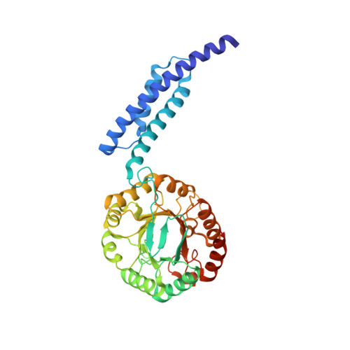
Interdomain Conformational Changes Provide Allosteric Regulation en Route to Chorismate.

Nazmi, A.R.Lang, E.J.M.Bai, Y.Allison, T.M.Othman, M.H.Panjikar, S.Arcus, V.L.Parker, E.J.

(2016) J Biological Chem 291: 21836-21847

  • DOI: https://doi.org/10.1074/jbc.M116.741637
  • Primary Citation Related Structures: 
    5J6F

  • PubMed Abstract: 

    Multifunctional proteins play a variety of roles in metabolism. Here, we examine the catalytic function of the combined 3-deoxy-d- arabino heptulosonate-7-phosphate synthase (DAH7PS) and chorismate mutase (CM) from Geobacillus sp. DAH7PS operates at the start of the biosynthetic pathway for aromatic metabolites, whereas CM operates in a dedicated branch of the pathway for the biosynthesis of amino acids tyrosine and phenylalanine. In line with sequence predictions, the two catalytic functions are located in distinct domains, and these two activities can be separated and retain functionality. For the full-length protein, prephenate, the product of the CM reaction, acts as an allosteric inhibitor for the DAH7PS. The crystal structure of the full-length protein with prephenate bound and the accompanying small angle x-ray scattering data reveal the molecular mechanism of the allostery. Prephenate binding results in the tighter association between the dimeric CM domains and the tetrameric DAH7PS, occluding the active site and therefore disrupting DAH7PS function. Acquisition of a physical gating mechanism to control catalytic function through gene fusion appears to be a general mechanism for providing allostery for this enzyme.


  • Organizational Affiliation
    • From the Biomolecular Interaction Centre and Department of Chemistry, University of Canterbury, P. O. Box 4800, Christchurch 8140, New Zealand.

Macromolecule Content 

  • Total Structure Weight: 83.82 kDa 
  • Atom Count: 5,420 
  • Modeled Residue Count: 692 
  • Deposited Residue Count: 736 
  • Unique protein chains: 1

Macromolecules

Find similar proteins by:|  3D Structure
Entity ID: 1
MoleculeChains  Sequence LengthOrganismDetailsImage
3-deoxy-D-arabino-heptulosonate 7-phosphate synthase, chorismate mutase-isozyme 3
A, B
368Geobacillus sp. GHH01Mutation(s): 0 
Gene Names: aroAGHH_c28860
EC: 2.5.1.54 (PDB Primary Data), 5.4.99.5 (PDB Primary Data)
UniProt
Find proteins for A0ACD6B8N4 (Geobacillus sp. GHH01)
Explore A0ACD6B8N4 
Go to UniProtKB:  A0ACD6B8N4
Entity Groups
Sequence Clusters30% Identity50% Identity70% Identity90% Identity95% Identity100% Identity
UniProt GroupA0ACD6B8N4
Sequence Annotations
Expand
Reference Sequence

Small Molecules

Ligands 3 Unique
IDChains Name / Formula / InChI Key2D Diagram3D Interactions
PRE

Query on PRE


Download:Ideal Coordinates CCD File
F [auth A],
J [auth B]
PREPHENIC ACID
C10 H10 O6
FPWMCUPFBRFMLH-XGAOUMNUSA-N
SO4

Query on SO4


Download:Ideal Coordinates CCD File
D [auth A],
E [auth A],
H [auth B],
I [auth B]
SULFATE ION
O4 S
QAOWNCQODCNURD-UHFFFAOYSA-L
MN

Query on MN


Download:Ideal Coordinates CCD File
C [auth A],
G [auth B]
MANGANESE (II) ION
Mn
WAEMQWOKJMHJLA-UHFFFAOYSA-N

Experimental Data & Validation

Experimental Data

  • Method: X-RAY DIFFRACTION
  • Resolution: 2.75 Å
  • R-Value Free:  0.235 (Depositor), 0.232 (DCC) 
  • R-Value Work:  0.182 (Depositor), 0.186 (DCC) 
  • R-Value Observed: 0.185 (Depositor) 
Space Group: P 64
Unit Cell:
Length ( Å )Angle ( ˚ )
a = 95.438α = 90
b = 95.438β = 90
c = 167.452γ = 120
Software Package:
Software NamePurpose
REFMACrefinement
Aimlessdata scaling
PHASERphasing
PDB_EXTRACTdata extraction
XDSdata reduction

Structure Validation

View Full Validation Report


Ligand Structure Quality Assessment 


Entry History 

Deposition Data

Revision History  (Full details and data files)

  • Version 1.0: 2016-09-07
    Type: Initial release
  • Version 1.1: 2017-09-27
    Changes: Data collection, Database references, Derived calculations
  • Version 1.2: 2023-09-27
    Changes: Data collection, Database references, Refinement description